

17 July 2026
Zebra shark eggs: ultrasound reveals the embryo without opening the case
Ultrasound monitoring of 120 zebra shark eggs clarifies when embryos and abnormalities may be detected, while showing why the images must remain cautious decision-support tools.
- Sector
- Public aquariums
- Themes
- ReproductionDiagnostics
- Animal groups
- FishElasmobranchs
- Content type
- Scientific news
Monitoring a shark inside its egg case presents a recurring challenge for aquariums. Teams need to distinguish a fertile egg from a non-viable one, recognise deterioration and anticipate hatching without disrupting development. Candling can provide useful information in some species, but the dark, tough egg case of the zebra shark (Stegostoma tigrinum) restricts direct observation. A study at the Aquarium of the Pacific shows that ultrasonography can reveal the embryo substantially earlier, using a submerged probe and without opening the case.
The findings are valuable, but they require careful interpretation. Of 120 embryonated eggs monitored between 2021 and 2023, only one produced a live hatchling. Most embryos with informative genetic results originated through parthenogenesis, meaning development occurred without paternal contribution. The research therefore describes a monitoring technique and prognostic signals in a highly unusual population. It does not establish a universal developmental calendar or an automatic rule for removing eggs.
A submerged probe and a consistently oriented egg case
The four adult females lived in two exhibits at the Aquarium of the Pacific, with seawater maintained between 23.8 and 25°C. Staff collected eggs daily, recorded laying dates and incubated them horizontally in continuously moving water at the same temperature range. That traceability was fundamental: without a laying date and temperature record, individual scans lose much of their comparative value.
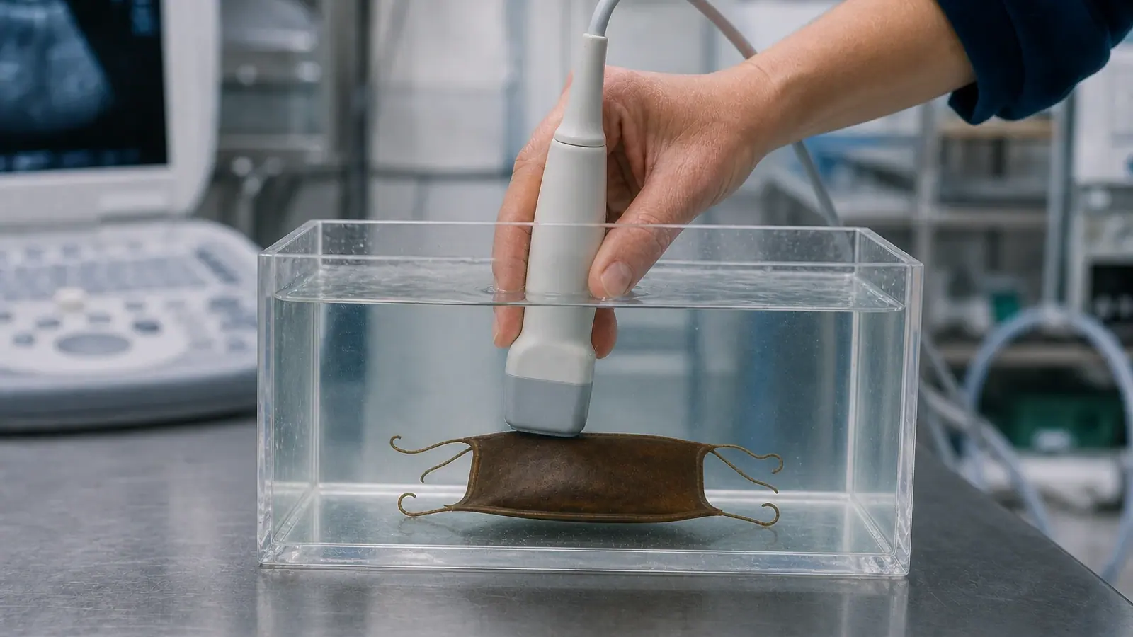
Following an initial 28–32 days without disturbance, most eggs underwent weekly scanning. Each egg case was transferred to a four-litre acrylic container filled with seawater while its orientation was maintained. The 15–6 MHz linear transducer was also placed in the water. Seawater provided acoustic coupling between probe and egg case, so no gel was applied to the case itself. Before the case naturally opened, the egg was exposed to air only briefly during transfer; after opening, transfers took place fully underwater.
This technique matters as much as the machine. Training material that depicts a probe scanning the case in air would misrepresent the procedure. The egg should remain immersed, supported and consistently oriented while transverse and sagittal views cover its entire length. The purpose is not to compress the case, but to obtain repeatable sequences with minimal handling.
Embryos could be seen from day eight in the earliest scans
Thirteen eggs were scanned weekly from early incubation. In this subset, the earliest embryo was detected at eight days, while the mean first observation occurred at 18 ± 3 days. Across all 120 eggs — 107 of which received their first scan only after three to five weeks — initial detection was around 30 days. The latest first detection occurred at 47 days.
This difference does not necessarily reflect biological variation; it is strongly shaped by examination timing. Presenting eight days as a guaranteed detection point would therefore be misleading. The practical finding is that ultrasound can reveal an embryo before it becomes readily visible by candling. Compared with historical data from the same institution, detection occurred at around 30 days with ultrasound rather than approximately 45 days with candling.
The smallest measured embryo was 0.18 cm long at 23 days. Total length generally remained measurable until days 64–70. Beyond that period, animal position and the ultrasound field of view reduced accuracy, particularly once embryos exceeded roughly 8 cm. The role of the scan then changes: rather than producing a simple growth curve, it helps document presence, movement, visible anatomy and the condition of the perivitelline environment.
Common abnormalities, but no absolute diagnosis
Ultrasound identified at least one abnormality in 84 of the 120 embryos, or 70%. The most frequent was a bent or coiled tail, recorded in 65 eggs. Vesicles on the yolk stalk were noted in 41 cases and altered head shape in six; nearly half of the abnormal embryos displayed more than one finding. Abnormalities were first detected at a mean of 43 ± 15 days, and none of those observed resolved during monitoring.
Another warning sign was flocculent material developing in the fluid around the embryo. It appeared in 41 eggs, representing 34% of the sample, at a mean of 45 ± 13 days. Every affected embryo subsequently died, on average 13 ± 16 days later. This association makes flocculent material a relevant alert in this dataset, but not yet a validated rule across other species, temperatures or breeding populations.
Conversely, the absence of a visible abnormality did not indicate a good prognosis. Among embryos with no detected ultrasound abnormality, cumulative mortality rose from 20% to 91.4% between days 35 and 56. The authors interpret this seemingly paradoxical pattern as an observation limit: some embryos probably died before reaching the developmental stage at which a malformation would have become visible.
Parthenogenesis demands a particularly cautious reading
Genetic samples were available from 65 embryos. Seventeen results were inconclusive, while 48 confirmed parthenogenetic origin, with at least one egg from each of the four females affected. No sexually produced embryos were available to establish a normal comparison timeline.
Only one of the 120 eggs hatched, a rate of 0.83%. Staff manually assisted hatching on day 205, about 45 days after the expected date, and genetic testing also confirmed that hatchling as a parthenote. Separate work involving aquarium-maintained zebra sharks has associated parthenogenesis with reduced growth and shorter survival compared with sexually produced offspring. That background is likely relevant to the high mortality observed here, but it does not allow each abnormality to be attributed to parthenogenesis.
Ultrasound should therefore not be used alone to classify an egg as infertile, doomed or ready for disposal. At the study temperature, persistent absence of an embryo after 47 days — approximately seven weeks — may strengthen suspicion of infertility or very early death. This benchmark remains specific to the species, facility and protocol. It must be interpreted alongside laying date, temperature, case appearance and changes across consecutive scans.
Building a useful aquarium protocol
For public aquariums, the main opportunity lies in standardisation. Each egg should have a unique identifier, collection date, incubation position and temperature history. Scans are most useful when performed by a trained operator using the same orientation, documented settings and archived images. Embryonic movement, measurable length, yolk appearance, perivitelline fluid and abnormalities should be described without inferring more than the images support.
Welfare and biosecurity remain central: keep transfers brief, use appropriate seawater, maintain immersion, clean equipment between groups and stop handling if the egg case resists or deteriorates. Any irreversible decision should involve the animal-care team and attending veterinarian, with reproductive or genetic specialists consulted where needed. Vetofish can help design the workflow, develop cautious interpretation standards and integrate findings into reproductive and veterinary records.
The study offers more than a new view of an embryo. It shows how a non-invasive technique can generate a usable longitudinal record. Its greatest value is not certainty about outcome, but earlier documentation of development and better-informed decisions that keep scientific limitations visible.
References
- Adams L, Wyffels JT, Goodwin B, Munson R, LeBorgne L, Feldheim KA, Lyons K. “Monitoring egg fertility, embryonic morbidity, and mortality in an oviparous elasmobranch using ultrasonography.” Frontiers in Veterinary Science. 2024;11:1410377. doi:10.3389/fvets.2024.1410377.
- Musa SM, Czachur MV, Shiels HA. “Oviparous elasmobranch development inside the egg case in 7 key stages.” PLOS ONE. 2018;13(11):e0206984. doi:10.1371/journal.pone.0206984.
- Adams L, Lyons K, Monday J, Larkin E, Wyffels J. “Costs of parthenogenesis on growth and longevity in ex situ zebra sharks Stegostoma tigrinum.” Endangered Species Research. 2023;50:81–91. doi:10.3354/esr01224.