

25 July 2026
Zebrafish: more new neurons do not guarantee better recovery
After spinal cord injury, disrupting sema4ab doubles progenitor and neuron production in zebrafish but reduces axon regrowth and swimming recovery.
- Sector
- Research facilities
- Content type
- Scientific news
- Keywords
- HistopathologyEthogramStress
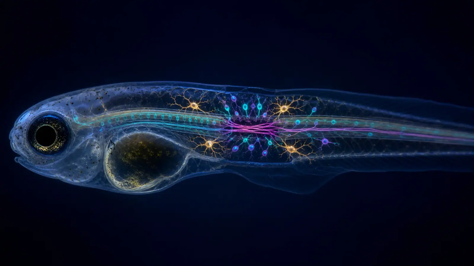
Zebrafish can generate new neurons and restore spinal connections after injury. A study in PLOS Biology shows, however, that maximising new-neuron production does not necessarily improve recovery. When sema4ab function was disrupted, newly generated progenitors and neurons doubled, yet axon regrowth and recovery of swimming were reduced. Repair depends on coordination, not on one cellular count.
A protein produced mainly by reactive microglia
Alberto Docampo-Seara and colleagues investigated Sema4ab, a semaphorin expressed mainly by lesion-reactive microglia. Microglia are resident immune cells of the central nervous system. After injury, they contribute to clearance, inflammation and signalling between neural progenitors, blood-derived macrophages and fibroblasts.
The authors combined single-cell RNA sequencing, imaging, proliferation labelling, genetic disruption and functional assessment in zebrafish larvae. This approach links gene expression to changes in cell populations and then to an observable outcome: swimming.
The model is powerful but specific. It uses a standardised experimental lesion in larvae over a defined observation window. It does not reproduce every adult spinal injury or the non-regenerative environment of a mammalian spinal cord.
Two routes that restrain neurogenesis
After sema4ab disruption, the researchers found approximately twice as many newly generated progenitor cells and neurons. One pathway may be direct. Neural progenitors express the Plexin-B1a and Plexin-B1b receptors, and disrupting those receptors also increased regenerative neurogenesis. Sema4ab may therefore deliver a signal that limits progenitor proliferation or differentiation.
A second route operates through the inflammatory environment. Without sema4ab, microglial activation state and cytokine expression changed. Fibroblasts at the lesion site then increased tgfb3 expression. In the reported experiments, Tgfb3 strongly promoted regenerative neurogenesis.
These relationships form a network rather than a single linear chain. The team did not identify the precise signal connecting Sema4ab-deficient microglia to fibroblasts. Disruption of candidate receptors and selected cytokine pathways did not reproduce every expected step. The indirect mechanism therefore remains partly unresolved.
More neurogenesis, poorer recovery
The most important result initially appears paradoxical. Disrupting sema4ab strongly increased neuron formation but attenuated axon regrowth and swimming recovery. Sema4ab therefore has different effects on several repair components: it restrains neurogenesis while supporting axonal growth and the measured functional outcome.
Successful repair depends on more than neuron number. Cells must acquire an appropriate identity, occupy the right location, extend axons, establish suitable connections and integrate into a circuit. Excessive or mistimed production could consume resources, alter inflammation or disturb tissue organisation. The study discusses these possibilities without establishing which one explains the functional decline.
This changes how a cellular “rescue” should be interpreted. Doubling a neurogenesis marker may appear favourable, but function must remain a decisive endpoint, accompanied by analysis of connectivity and the quality of generated cells.
A mouse comparison, not yet a treatment
The authors reanalysed single-cell data from mouse spinal injury. They found a compatible arrangement: Sema4A expression in microglia, Plexin-B1 in progenitors and Tgfb3 in fibroblasts. This suggests that elements of the network are conserved across vertebrates.
It does not demonstrate that inhibiting Sema4A would restore a mammalian spinal cord. Concentrations, timing, receptors and scar environments differ. More importantly, the zebrafish study itself shows that an intervention favouring neurogenesis can impair axon growth and function. Any translational strategy would have to balance several outcomes rather than optimise a single cellular measure.
Future experiments can define when Sema4ab is beneficial or restrictive, identify the microglia-to-fibroblast signal and characterise the additional neurons. Cell-type-specific and time-restricted manipulations should help separate direct signalling from secondary inflammatory effects.
Implications for experimental-model quality
For zebrafish facilities, the work highlights the need to standardise health status, developmental stage, temperature, lesion timing and imaging conditions. Inflammation and regeneration are context-sensitive. Subclinical infection, microbiome differences or repeated handling can add variation to immune and locomotor profiles.
Swimming assessment needs a defined timeline and appropriate controls. Reduced activity may reflect motor impairment, but it can also arise from a general manipulation effect or developmental difference. Convergence between anatomy, cell markers, axonal measures and behaviour strengthens interpretation.
Experimental unit and blinding also matter. Larvae from the same clutch or vessel are not necessarily independent in every analysis. Reporting allocation, exclusion criteria and image-analysis thresholds supports reproducibility, particularly where effects are followed across multiple levels.
Conclusion: coordination matters more than quantity
Sema4ab is not simply a harmful brake. It limits new-neuron production while contributing to conditions that support axon regrowth and swimming recovery. Removing it yields more neurogenesis but a poorer functional result.
Vetofish can support zebrafish platforms with health surveillance, procedural standardisation and joint interpretation of welfare and functional endpoints. The study is a reminder that cell counting is necessary in regeneration research but never sufficient: timing, connectivity and whole-animal behaviour remain essential.
References
- Docampo-Seara, A., Cosacak, M. I., Heilemann, K. et al. (2026). “The microglia-derived protein Sema4ab attenuates regenerative neurogenesis after spinal cord injury in zebrafish.” PLOS Biology, 24(6), e3003865. https://doi.org/10.1371/journal.pbio.3003865
- Cavone, L., McCann, T., Drake, L. K. et al. (2021). “A unique macrophage subpopulation signals directly to progenitor cells to promote regenerative neurogenesis in the zebrafish spinal cord.” Developmental Cell, 56(11), 1617–1630.e6. https://doi.org/10.1016/j.devcel.2021.04.031