

17 July 2026
Tumours in public-aquarium fish: observe, sample and diagnose without jumping to conclusions
A retrospective study describes 63 tumours in 62 fish from 37 species and shows why a visible mass requires longitudinal records, purposeful imaging and histological confirmation.
- Sector
- Public aquariums
- Themes
- DiagnosticsDiseases
- Animal groups
- FishElasmobranchs
- Content type
- Scientific news
A skin mass, a distorted eye or progressive abdominal enlargement cannot identify a tumour by appearance alone. In a public aquarium, teams must protect the fish’s welfare while obtaining enough evidence to guide treatment, population surveillance and quality-of-life decisions. A 2024 retrospective study in the Journal of Veterinary Diagnostic Investigation reviewed neoplasia diagnosed at Vancouver Aquarium from July 2005 to October 2021.
The authors identified 62 fish from 37 species that met their inclusion criteria. One fish had two tumours, giving 63 neoplasms and 32 diagnostic types. Ten were benign and 53 malignant. These numbers demonstrate diagnostic diversity, but they are not a cancer prevalence estimate: the study did not screen every fish in the collection or provide the full population denominator across the 16-year period.
Evidence drawn from records, biopsies and postmortems
Sarah Wright and colleagues reviewed medical records, biopsy reports and postmortem findings from fish diagnosed with neoplasia before or after death. Samples were fixed in 10% neutral-buffered formalin and examined histologically. Five ACVP-certified veterinary pathologists contributed over the study period, and at least two pathologists reviewed every case to reach a consensus diagnosis.
The case series included 60 teleosts and two elasmobranchs. Thirty-four fish represented 18 tropical freshwater species, eleven represented nine tropical marine species, and seventeen represented ten cold-water marine species. The records included 14 biopsy reports from eleven fish and 54 postmortem reports. Three fish had both.
The two routes answer different questions. A biopsy may establish a diagnosis in a living animal and support a treatment decision. A postmortem examination can reveal internal distribution, clarify the primary site and create knowledge that improves care for the rest of the collection.
Malignancy dominated the diagnosed case series
Among the 63 tumours, the authors classified 24 as epithelial, twenty as neuroectodermal or neural-crest derived, twelve as mesenchymal, four as lymphomas and three as germ-cell tumours. Neuronal embryonal tumours and papillomas were most frequent, with seven cases each. Thyroid carcinomas and malignant peripheral nerve-sheath tumours followed with six each. Four lymphomas and four iridophoromas were also reported.
Skin was the most common site of origin. In tropical freshwater fish, 29 of 35 tumours were malignant. All eleven tropical marine tumours were malignant, as were thirteen of seventeen tumours in cold-water marine fish. The authors suggested that mesenchymal neoplasia might be more common in cold-water marine fish than in the two tropical groups.
This pattern must not be converted into an individual risk estimate. The study only contains diagnosed cases and is affected by submission bias. Some fish underwent histology while others did not, including carcasses rejected because of advanced autolysis. The year with most recorded cases probably reflected more histology submissions rather than a proven increase in incidence.
A mass is not automatically a neoplasm
In a living fish, an external swelling may also be an abscess, granuloma, cyst, inflammatory reaction, hyperplasia or parasitic proliferation. Internal masses may present as asymmetry, reduced appetite, buoyancy change or loss of body condition, all of which are nonspecific.
The first diagnostic layer is a timeline: onset, growth rate, location, pigmentation, ulceration, bleeding, locomotor interference, food intake and social behaviour. Photographs taken from a repeatable angle with a scale provide more reliable evidence of change than memory or casual visual comparison.
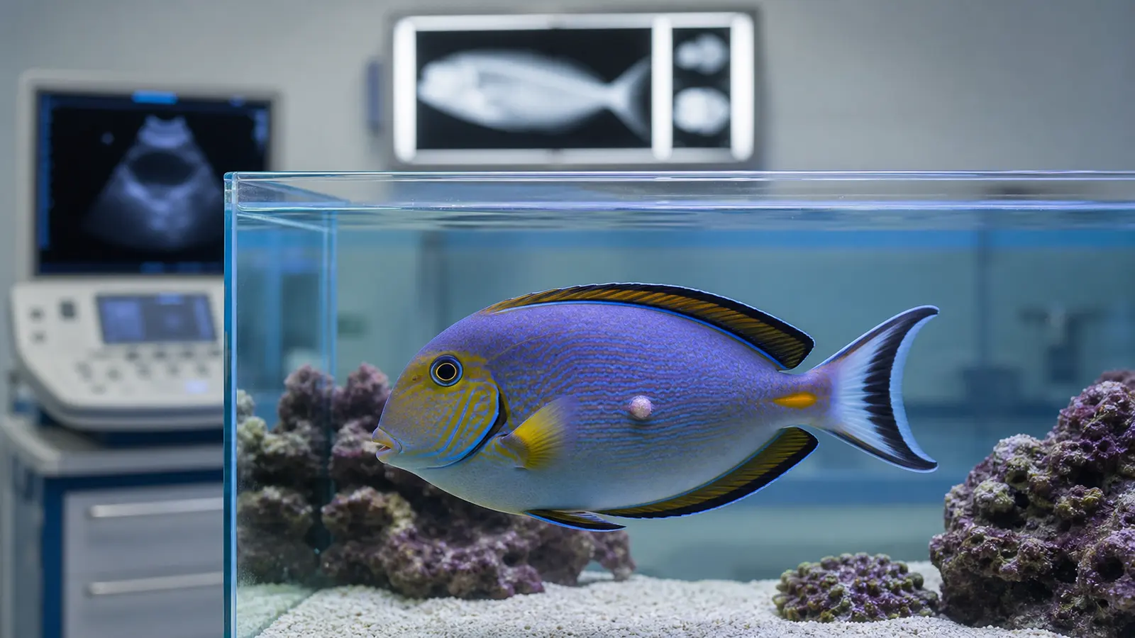
Ultrasonography can help distinguish solid from fluid-filled coelomic lesions, assess relationships with organs and plan sampling. Radiography can evaluate bone involvement, mineralisation and some organ displacement. Imaging guides the next step but does not replace histology. Its value depends on species, body size, lesion location and the welfare cost of capture and anaesthesia.
Preserve diagnostic value when sampling
A useful biopsy needs adequate depth and, when safe, the transition between abnormal and apparently normal tissue. A superficial or entirely necrotic fragment may prevent classification. The choice between incisional biopsy, excision and monitoring depends on accessibility, suspected infiltration, local function and overall health.
The whole procedure should be planned before capture: equipment, anaesthesia, oxygenation, haemostasis, recovery and the destination of every sample. Tissue for histology should enter a suitable volume of 10% neutral-buffered formalin promptly. If infection remains a differential, separate aseptic samples for microbiology must be collected before the tissue contacts fixative.
At postmortem examination, gross description, photographs and samples from apparently normal organs are as important as the mass itself. Several tumours in the Canadian series involved deep or multiple tissues. Sampling only the most visible lesion may therefore obscure origin and distribution.
Connect diagnosis with quality of life
A malignant diagnosis does not make every clinical decision automatic. Species, estimated age, rate of progression, likely discomfort, feeding, swimming, social interaction, treatment feasibility and the burden of repeated capture all matter. A small stable mass may justify structured observation, whereas an ulcerated, invasive or function-limiting lesion may require earlier intervention.
Monitoring should use predefined criteria: mass growth, body condition, intake, ventilation, swimming, habitat use and secondary lesions. Agreed intervention thresholds reduce the risk that gradual deterioration becomes normalised by a team that sees the fish every day.
Stephen Smith’s review of welfare in public aquariums recommends combining behavioural, performance, physiological and environmental indicators. Applied to a fish with a mass, that framework prevents the lesion’s appearance from becoming the only measure of welfare.
Build a collection-level health record
The retrospective study demonstrates the value of consistent terminology, longitudinal records and postmortem examination whenever carcass quality permits. An internal database linking species, estimated age, provenance, habitat, exposures, histological diagnosis and outcome can reveal clusters while guarding against premature claims of a common cause.
Vetofish can support public aquariums with mass triage, biopsy planning, pathology interpretation and quality-of-life scoring. The practical sequence is straightforward: observe consistently, use imaging to answer a defined question, and preserve a diagnostic sample. Histological confirmation turns a visible abnormality into medical information that can be shared and compared.
References
- Wright SE, Pawlik M, Snyman HN, Haulena M. “Review of neoplasia in fish at a large display aquarium, 2005–2021.” Journal of Veterinary Diagnostic Investigation. 2024;36(3):362–367. doi:10.1177/10406387241241344.
- Smith SA. “Fish Welfare in Public Aquariums and Zoological Collections.” Animals. 2023;13:2548. doi:10.3390/ani13162548.