

17 July 2026
Mycobacterium haemophilum in zebrafish: diagnosing beyond appearances
This fastidious mycobacterium can cause systemic disease in laboratory zebrafish and persist in biofilms, calling for integrated fish and environmental surveillance.
- Sector
- Research
- Themes
- DiseasesDiagnostics
- Content type
- Disease profile
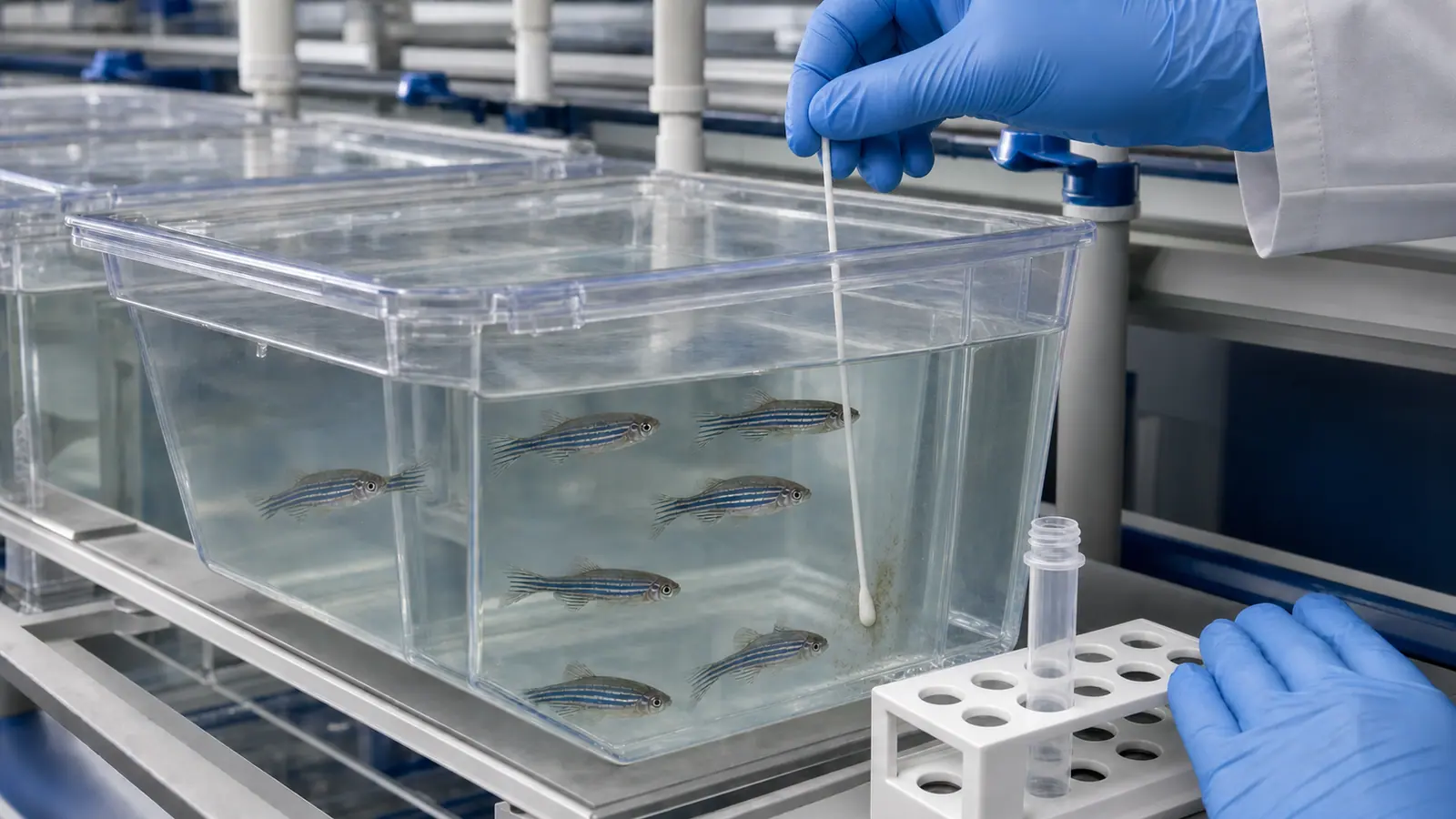
Chronic mortality, skin ulcers or loss of condition in a zebrafish facility may initially look nonspecific. Mycobacterium haemophilum demands a wider view. This nontuberculous mycobacterium can cause severe systemic infection in Danio rerio, while its unusual growth requirements make routine culture less reliable unless the laboratory is alerted. The organism has also been detected in biofilms within recirculating systems, making the aquatic environment part of the diagnostic investigation rather than a passive backdrop.
Two documented outbreaks in research facilities define the problem. In 2005, Whipps and colleagues isolated M. haemophilum during substantial losses and reproduced the disease experimentally. A later outbreak at the University of Glasgow affected a wild-origin colony and its offspring; after losing almost the entire population, the team described how it decontaminated and repopulated the system. Neither account provides a universal recipe. Together, however, they offer evidence for a structured approach to surveillance, diagnosis and management of a contaminated loop.
Systemic disease without a single defining presentation
In the 2007 investigation, infected fish developed granulomas and severe diffuse chronic inflammation. Bacteria occurred in multiple tissues, including the central nervous system, and experimental infection confirmed the pathogenicity of the isolate. Biofilm samples from the affected facility were PCR-positive for M. haemophilum, supporting the possibility that established biofilms can act as an environmental reservoir.
The Glasgow outbreak produced raised scales, ulcers, ascites, lethargy, emaciation and haemorrhage. At one sampling point, 80 fish in an F1 population of 1,400 had been quarantined with clinical signs. Histology, acid-fast staining, bacteriology and species-specific PCR were used to characterise the event. Evidence of infection was found in fish as well as in system water and biofilm.
Clinical examination alone can therefore neither identify the bacterial species nor exclude less visible circulation. Granulomatous lesions support a diagnosis of mycobacteriosis but do not resolve the organism to species. Conversely, a positive PCR detects a genetic target; on its own, it does not demonstrate tissue damage or establish that the organism explains every clinical case. Molecular and pathological findings answer different questions and are strongest when interpreted together.
Why conventional culture may miss the organism
The name haemophilum points towards an important diagnostic feature: growth requires an iron source such as hemin. In the qPCR development study by Meritet and colleagues, the reference isolate was grown on Middlebrook 7H10 agar supplemented with hemin for 28 days at 32 °C. The authors also note that some mycobacteria are difficult to grow and that biochemical identification can be uninformative.
A standard culture request that does not mention suspected M. haemophilum may therefore use unsuitable conditions. The diagnostic laboratory needs the host species, lesion pattern, fish origin, system temperature and prior treatment so it can select appropriate media, incubation temperature and duration. A negative result is meaningful only in relation to the tissue sampled, its preservation and the procedure actually performed.
Species-specific qPCR provides a faster identification route. The assays developed by Meritet and colleagues targeted the hsp65 gene of M. haemophilum, M. marinum and M. chelonae. All experimentally infected fresh-frozen fish assessed in the study were detected by the corresponding assays. Sensitivity was more moderate in formalin-fixed, paraffin-embedded material. For M. haemophilum, cores aimed at granulomas within the block performed better than whole-fish scrolls. Performance from fresh tissue should not be assumed for a fixed specimen.
Representative numbers and informative locations
Even a technically strong assay cannot rescue a narrow sampling plan. Marancik and colleagues compared subpopulations in a recirculating research facility with 1,000 tanks and approximately 30,000 fish. Randomly selecting 60 fish and sampling 53 sump fish detected a broader range of agents than smaller groups of 11 sentinels or 18 moribund or recently dead fish. In that facility, M. haemophilum was frequently identified in several groups but was not detected in the sentinel group.
This does not show that sentinels are universally ineffective. It shows that a small population assumed to be at risk may not be representative and that a negative result depends on accessible prevalence, exposure and sample size. Under the study’s assumptions, 60 fish provided 95% confidence of detecting an agent at a prevalence of at least 5.54%; for 11 sentinels, the corresponding threshold was 26.5%. Those figures belong to the specific design and should not be transferred without accounting for assay sensitivity, pooling and facility structure.
A coherent investigation may combine recently affected fish, a representative population sample, histopathology, species-specific PCR and environmental swabs from sites where organic material accumulates. Each hydraulic loop also deserves separate consideration. In the retrospective part of Marancik’s study, a pathogen found in one system was sometimes absent from another system in the same facility.
Turning a positive result into a health decision
Management depends on the distribution of infection, the value of affected lines, options for rederivation, hydraulic design and research objectives. At Glasgow, the remaining colony was culled before two 340-litre units and six 300-litre aquaria underwent a staged process involving dismantling, physical cleaning, chlorination and secondary use of Virkon Aquatic. Six months after disinfection, tested fish, water and biofilm samples were negative for M. haemophilum. Further environmental testing took place about one year after the procedure, and the replacement colonies did not show comparable disease-associated mortality during the first year.
These are valuable results from one intervention after animal removal, not instructions to improvise chemical treatment. Concentration, contact time, material compatibility, neutralisation, occupational safety and biofilter restart must be validated with manufacturers, the facility veterinarian, technical managers and the diagnostic laboratory. Applying these disinfectants to occupied tanks or borrowing a concentration without checking the whole protocol would be unsafe.
Post-decontamination surveillance should be designed before restocking. Sampling points, timing, acceptance criteria and the response to discordant findings all need to be explicit. Movement records, separate quarantine equipment and controls on water transfer reduce the likelihood of reintroduction. These measures also support science: a subclinical mycobacterial infection may change inflammation, immunity, metabolism or survival and thereby become an unmeasured source of experimental variation.
Protecting animals, evidence and staff
M. haemophilum is recognised as a cause of human infection, especially in immunocompromised people. The zebrafish studies do not, however, demonstrate transmission from a laboratory colony to staff. A proportionate response is to include the organism in local risk assessment: suitable gloves, protection of broken skin, control of splashes and aerosols during cleaning, and access to occupational-health advice after a concerning exposure.
The goal is not indiscriminate testing. It is to connect clinical observations, pathology, molecular detection and system surveillance, then interpret each result within its limitations. Vetofish can support sampling-plan design, laboratory selection, health-risk assessment, biosecurity and verification before and after restocking. With M. haemophilum, a robust diagnosis protects zebrafish welfare, experimental reproducibility and the people who maintain the colony.
References
- Rácz A, Dwyer T, Killen SS. “Overview of a Disease Outbreak and Introduction of a Step-by-Step Protocol for the Eradication of Mycobacterium haemophilum in a Zebrafish System.” Zebrafish. 2019;16(1):77-86. Published online 24 October 2018. doi:10.1089/zeb.2018.1628.
- Whipps CM, Dougan ST, Kent ML. “Mycobacterium haemophilum infections of zebrafish (Danio rerio) in research facilities.” FEMS Microbiology Letters. 2007;270(1):21-26. doi:10.1111/j.1574-6968.2007.00671.x.
- Meritet DM, Mulrooney DM, Kent ML, Löhr CV. “Development of Quantitative Real-Time PCR Assays for Postmortem Detection of Mycobacterium spp. Common in Zebrafish (Danio rerio) Research Colonies.” Comparative Medicine. 2017;67(2):131-141. PMCID: PMC5361037.
- Marancik D, Collins J, Afema J, Lawrence C. “Exploring the advantages and limitations of sampling methods commonly used in research facilities for zebrafish health inspections.” Laboratory Animals. 2020;54(4):373-385. doi:10.1177/0023677219864616.
- Mocho J-P, Collymore C, Farmer SC, Leguay E, Murray KN, Pereira N. “FELASA-AALAS Recommendations for Monitoring and Reporting of Laboratory Fish Diseases and Health Status, with an Emphasis on Zebrafish (Danio rerio).” Comparative Medicine. 2022;72(3):127-148. doi:10.30802/AALAS-CM-22-000034.