

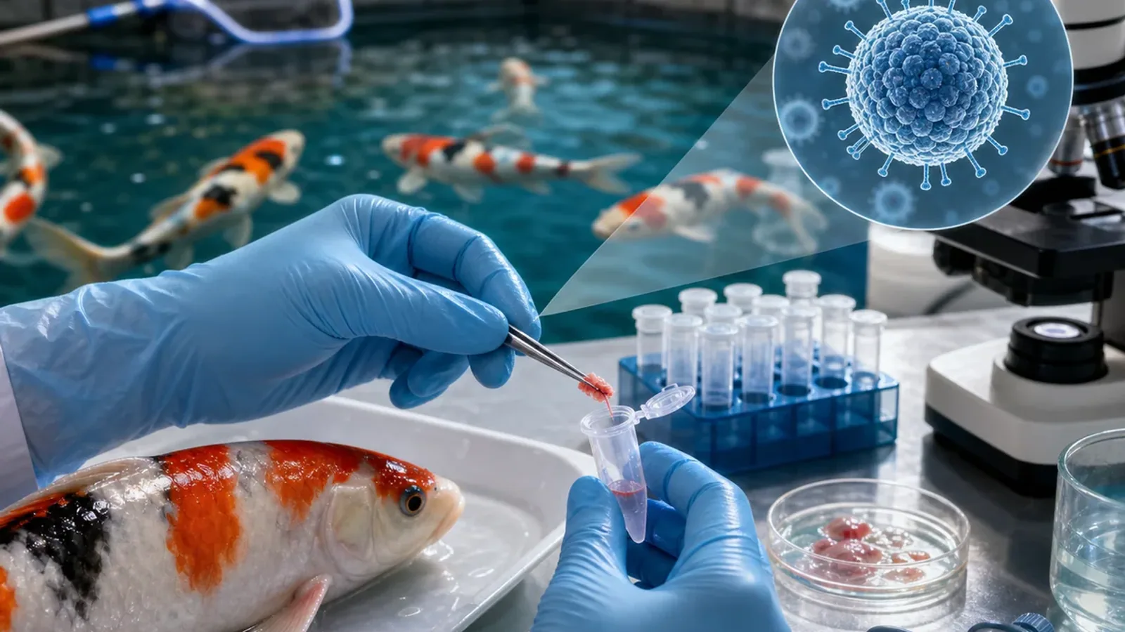
How is KHV (CyHV-3) detected in carp?


KHV detection relies mainly on molecular methods, including real-time PCR, performed by a competent laboratory. The laboratory and veterinarian select the method for the actual purpose: confirming clinical disease, surveying an apparently healthy stock or investigating past exposure.
Which fish and samples should be selected?
During an outbreak, moribund, abnormal or very recently dead fish usually offer the best detection probability. In clinically affected fish, gill, kidney and spleen are among the recommended tissues. A different plan is needed for apparently healthy carriers and should be agreed with the laboratory before sampling.
The WOAH manual states that the non-lethal methods studied so far do not have sufficient formal validation to be recommended as a general KHV detection method. A simple swab should therefore not be presented as proof that a fish is free from infection.
How should a result be interpreted?
A positive PCR detects a viral DNA sequence; confirmation and interpretation depend on the assay, controls and context. A negative PCR only means that the target was not detected in the samples tested. It may fail to exclude latent infection, a low viral load, unsuitable timing or an inappropriate sample.
Serology looks for an immune response and may support some investigations, but it cannot by itself prove that a fish is shedding virus or that a stock is disease-free.