

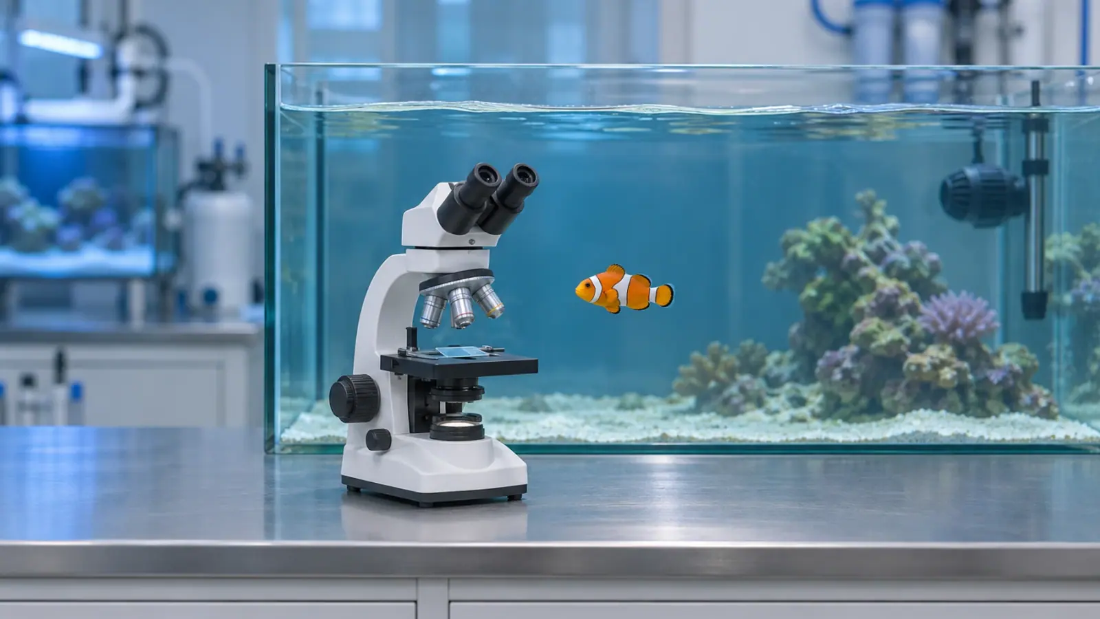
How can you tell Cryptocaryon irritans from Amyloodinium ocellatum?
A fresh wet mount can distinguish the two parasites. Cryptocaryon irritans trophonts have cilia and often roll or rotate under the microscope. Amyloodinium ocellatum trophonts are usually still and attached to tissue. Distinct white spots suggest marine white spot disease, while a fine haze can suggest marine velvet. Either parasite can affect the gills of a fish whose skin looks clear.
When should the gills be examined?
If several fish are breathing rapidly, gathering by a water inlet or going off feed, check dissolved oxygen and water circulation and arrange a prompt veterinary examination. Amyloodiniosis can progress quickly. In the European sea bass studied by Massimo and colleagues, the gills and the lining of the mouth and throat were the main sites affected. Some fish died with no obvious skin lesions. That finding shows what an external examination can miss in sea bass. Other species may show a different pattern.
Discrete spots on the skin or fins increase suspicion of Cryptocaryon. If the infestation mainly affects the gills, rapid breathing may come first or be the only visible sign. How quickly the fish deteriorate helps set the urgency. Water readings and samples help establish the cause.
What does the veterinarian look for on a wet mount?
The signs guide sampling. Spots on the skin or fins give the veterinarian a site to examine. Laboured breathing with clear skin makes the gills a priority. The veterinarian decides which fish to sample, taking into account how well they can tolerate handling.
Francis-Floyd and Floyd’s SRAC guide compares the feeding stages, known as trophonts. On a fresh wet mount, C. irritans can roll or move on or within the tissue. An A. ocellatum trophont is often brown, oval or pear-shaped and usually stays still. The two can overlap in shape and size. Look at cilia, movement and how the organism sits against the tissue together. A wet mount made with water of suitable salinity helps keep Amyloodinium trophonts attached to the sample.
A clear skin sample leaves open the possibility of a gill infestation. If fish continue to deteriorate, the veterinarian can examine other fish or ask the laboratory to confirm the identification. The next step depends on which tissue was examined, the condition of the fish and what else might be causing respiratory distress.
What can the team do while waiting for results?
Record which species and tanks are affected, when signs began, recent introductions and which tanks share water. Pause transfers and keep equipment within the affected system. Both parasites have stages that develop away from the fish. When the spots or haze disappear from one fish, the risk of reinfection in the tank can persist.
For surveillance across connected tanks, see how to detect Amyloodinium early in a marine aquarium. Vetofish’s testing and diagnosis service can help select samples and interpret the findings alongside the aquarium records.