

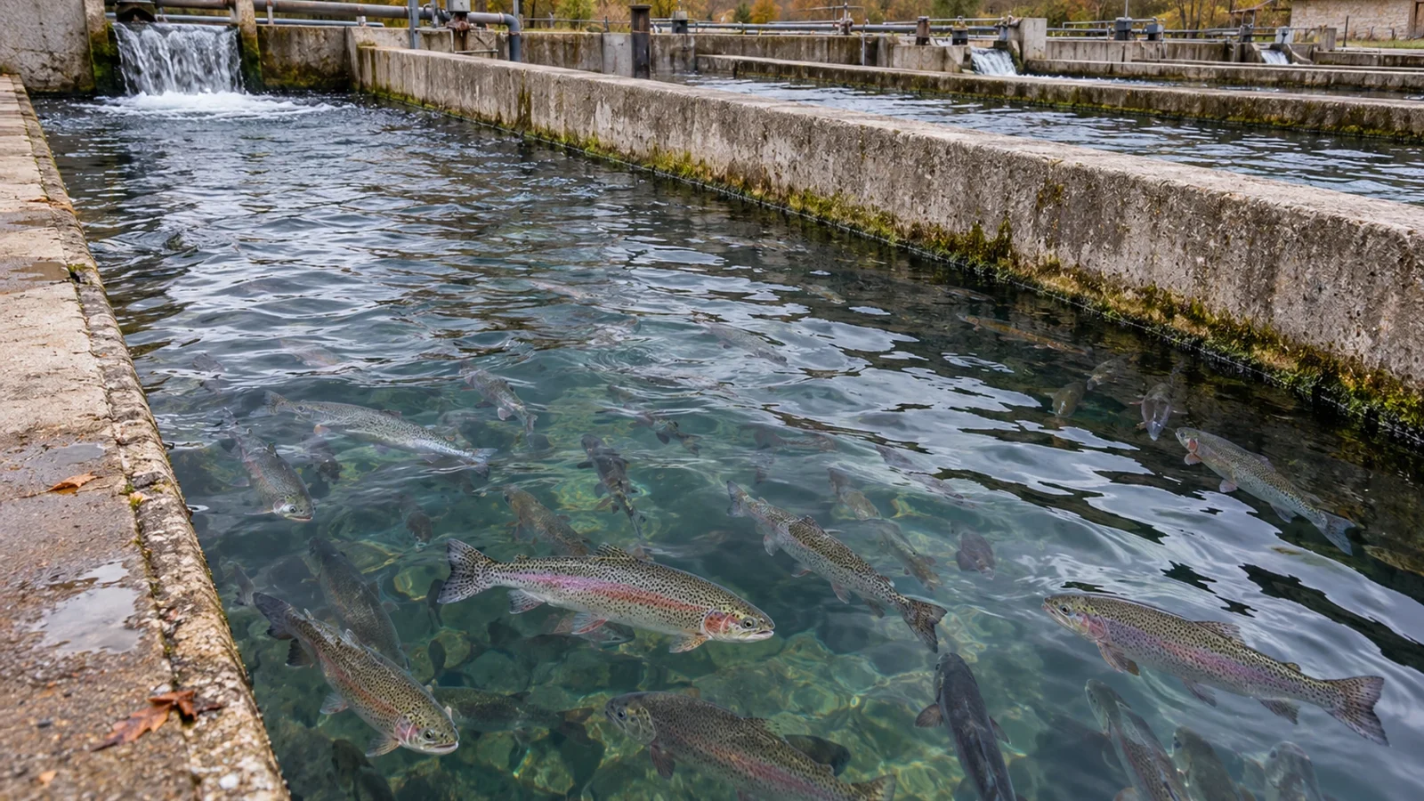
Injured trout: interpreting Saprolegnia DNA detection
Injured trout can carry more Saprolegnia parasitica DNA without visible cotton-like growth. A 2022 study helps separate pathogen detection from tissue disease and clinical risk, while providing no treatment threshold.
- Content type
- Practical guide
- Sector
- Aquaculture
- Theme
- DiseasesDiagnostics
A trout can have injured skin without the conspicuous cotton-like growth associated with saprolegniosis. Equally, finding Saprolegnia parasitica DNA on that skin does not establish that the organism caused the injury. These distinctions help a farm decide what to investigate and how to read the laboratory result.
Pavić and colleagues examined this problem in a 2022 study involving adult trout from Croatian farms. Their work developed a detection method and compared injured with apparently healthy fish. It did not establish a treatment trigger or test a clinical management programme. Its practical value lies in defining what a positive result can support.
The adult trout comparison
Skin swabs came from thirty adult trout sampled at four farms during winter 2018–2019. The sample included rainbow trout, Oncorhynchus mykiss, and brown trout, Salmo trutta. Seventeen fish had skin injuries and thirteen appeared healthy. The injured fish lacked gross signs of saprolegniosis at the time described.
These adults were already dead when sampled, having been collected during routine farm harvesting. This part of the research therefore did not validate a clinical surveillance procedure by following live fish after swabbing. The paper also examined eggs and environmental waters. Those samples answer related questions but are separate from the thirty adult fish.
The distinction between an injured fish and a confirmed disease case is central. A lesion is an observation requiring explanation. It does not identify an organism, establish tissue invasion or show which event occurred first. The comparison was designed to examine association with pathogen DNA load rather than resolve all these clinical questions.
What droplet digital PCR detects
Saprolegnia belongs to the oomycetes, filamentous organisms distinct from true fungi. The assay targeted a region of its DNA using droplet digital PCR, usually abbreviated to ddPCR. Dividing the reaction into droplets allows the amount of target material to be estimated from positive reactions.
The study expressed results as ITS target copies per quantity of total extracted DNA. That unit is not a direct count of viable organisms, a measurement of invaded tissue or a description of lesion area. The sampled surface, extraction process and other biological material on the swab also matter to interpretation.
The analytical assessment included closely related organisms. A small signal nevertheless occurred with one non-target strain. Specificity should therefore be understood within the tested conditions. The word molecular does not remove the need for appropriate controls, knowledge of the assay and interpretation by the laboratory.
More DNA on injured fish, without a disease threshold
Swabs from injured trout carried higher S. parasitica DNA loads than swabs from apparently healthy trout. One injured fish was negative. That exception is useful clinically: similar-looking injuries need not have identical microbial associations, and an injured surface cannot identify the responsible agent on its own.
The comparison did not determine whether the organism initiated the injury, colonised it afterwards or occurred alongside another process. Nor did it follow the adult fish through subsequent disease development. A copy number from this study cannot be converted into a universal threshold for saprolegniosis or a decision to medicate.
Build the sampling plan around the clinical question
A farm investigation needs a lesion description, location, time course and record of recent handling. The number affected, stock at risk and mortality history provide context that the laboratory cannot reconstruct from a tube. Dated photographs can help track change, while leaving diagnosis to an appropriate examination.
The veterinarian and laboratory can decide whether surface swabs, lesion margins, tissues or water address the question. Collection, transport and storage should suit the intended analysis. DNA detection, culture and histopathology may require different samples or preservation conditions, so the plan should be agreed before material is collected.
Where tissue invasion is the question, histopathology can complement surface detection. Results must still be connected to the same investigation. A positive swab from one fish and an unrelated lesion on another do not automatically demonstrate causation. Clear identities make it possible to assess whether different lines of evidence agree.
Put the skin findings back into farm history
Sorting, transfers, contact with equipment and changes in water conditions can be recorded alongside injuries. This is a way to develop and examine hypotheses, rather than prove that every lesion came from a particular operation. Repeated patterns are easier to assess when the sequence of events is preserved.
Sampling biosecurity matters too. Equipment moving between units may obscure where a signal originated. Fish, units, dates and operations need distinct identities so that an individual finding is not mistaken for a problem shared throughout the site. The objective is an interpretable investigation, rather than the largest possible collection of samples.
Negative findings should remain in the record. Sampling only severely affected animals gives a limited view of the stock. The veterinarian can help select relevant comparators and decide when an additional capture would provide useful information. Collecting more material without a defined question can add disturbance without resolving uncertainty.
Move from detection to a supported decision
The study gives a reason to investigate damaged skin even before visible cotton-like growth appears. It also keeps pathogen presence, tissue involvement and disease as separate questions. Decisions depend on the clinical picture and farm conditions, with a laboratory result interpreted in relation to its actual sample.
Vetofish diagnostic support can help define appropriate material and assess whether the findings agree. For aquaculture operations, the aim is to understand the injuries and the possible contribution of Saprolegnia before choosing proportionate health measures.


