

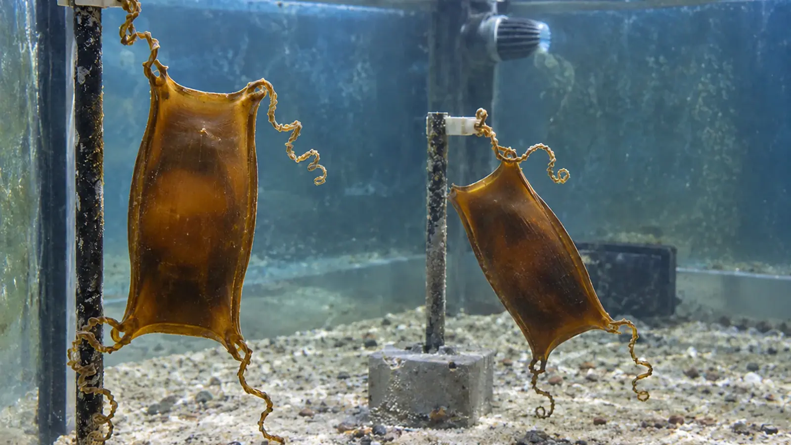
Catshark egg cases: follow development before hatching
A seven-stage key helps aquarium teams follow development inside greater spotted catshark egg cases. It provides observation markers, rather than a deadline for assisted opening.


- Content type
- Practical guide
- Sector
- Public aquariums
- Animal group
- Elasmobranchs
- Keywords
- ReproductionTemperatureOxygen
Follow the egg case as an individual
In the nursehound or greater spotted catshark, Scyliorhinus stellaris, the egg case protects the developing young and contributes to its relationship with the surrounding water. Seeing movement or a partly visible outline does not establish that the case should be opened. Aquarium staff need a record of developmental progress before they can interpret a suspected delay.
Musa, Czachur and Shiels published a seven-stage description in 2018, developed mainly in this species and compared with the small-spotted catshark, Scyliorhinus canicula. It offers observable developmental markers. It is not a clinical trial of assisted hatching and does not validate one intervention protocol for every egg-laying shark.
Each case therefore needs an individual identifier, confirmed species and laying date where known. Keep successive photographs with water-condition records. Without that history, a normal stage may look delayed, while a genuine change may be difficult to recognise. Staff should also record uncertainty when the contents cannot be seen clearly.
Describe visible changes rather than guessing age
The first stage has no embryo visible to the unaided eye, although important development is taking place. A small embryo then becomes visible on the yolk, followed by a longer tail. External gill filaments develop and later shorten. Fin folds disappear at a subsequent stage, while the animal increasingly resembles the hatchling form.
In the final stage, the external yolk sac has largely disappeared. Yolk supplies material and energy during development, but its apparent volume is not a direct measurement of remaining energy. Interpret the external sac alongside body development and the overall sequence, rather than treating any decrease as a signal to release the animal.
The useful contribution of the key is a shared vocabulary for biological progression. A monitoring record can identify the proposed stage, the features supporting it and those obscured by the case. An opaque case cannot support a confident stage assignment merely because its laying date is known. Likewise, no movement during one short observation is insufficient evidence that the embryo has died.
Put incubation time beside the rearing conditions
At 15 °C, the study reported mean development times to hatching of 27.25 weeks for S. stellaris and 24 weeks for S. canicula. Eight individuals of each species contributed to this comparison. These are observations from small groups under specified conditions, rather than a deadline beyond which staff should cut an egg case open.
Temperature records and changes in the holding system belong beside the photographs. Comparing an animal maintained under different conditions with one published mean can generate a misleading alert. The mean also conceals individual variation, even when species and conditions match. Progress through recognisable stages is more informative than a calendar alone.
The authors used a salinity of 35 ppt and dissolved oxygen above 95% saturation. Those values describe their experimental setting. They should not be copied as universal aquarium operating instructions. A local team needs species-specific husbandry information, reliable measurements and an understanding of the conditions around the cases themselves, supported by veterinary advice.
Preserve the early protective structures
The case and its internal jelly provide protection during early development. The study describes seawater slits opening during a biological transition as the plugs and jelly change. Prematurely imposing seawater exposure had adverse consequences in the S. stellaris observations. This finding matters when considering apparently simple alterations to a case.
Shaving its surface, cutting an end or removing a protective structure can change the conditions experienced by the embryo. Some procedures in the paper were used to describe anatomy, including examination of embryos removed from cases or euthanised. They are research methods, not instructions for the routine care of live eggs in a public aquarium.
The practical monitoring approach proposed here favours brief, comparable observations, limited handling and keeping the cases submerged. Candling is useful only when it provides interpretable information under suitable conditions. A photograph should be described according to what is visible, including limitations, rather than turned into a diagnosis because the outline looks unusual.
A slow exit is not an intervention threshold
The authors observed hatchlings rotating so that the head pointed towards the opening, then pushing against the case with the tail. Escape took a few minutes in some observed individuals and more than an hour in others. That variation prevents a stopwatch alone from defining an emergency.
When difficulty is suspected, bring together the developmental stage, recent observations, case condition and water measurements. A system change, a visible alteration or an inconsistent developmental sequence warrants veterinary assessment. The paper does not provide a validated threshold for assisted opening, and this account does not recommend routine incisions.
Before any intervention, identify the problem it is intended to solve and prepare subsequent care. The decision depends on species, stage and examination. Releasing an animal that still depends on embryonic structures can introduce a different problem from the one staff initially suspected. The aim is an informed response, supported by evidence from that individual.
Carry the history into hatchling care
The egg-case record should stay with the hatchling. Record the date and circumstances of emergence, external appearance, behaviour and initial observations in its receiving environment. Plan transfer and feeding around species and condition. Loss of the external yolk sac does not establish that all internal reserves have been exhausted.
The seven-stage key helps keepers and veterinarians discuss progress consistently. Its experimental timetable cannot replace examination or a coherent local record. A documented sequence gives the team a stronger basis for deciding whether continued observation or a clinical assessment is appropriate, and preserves useful information for future breeding work.
Develop the monitoring record with Vetofish
Vetofish can help design observation sheets, identify findings that require veterinary discussion and prepare proportionate intervention decisions. Our advice and support assists public aquarium teams with reproductive monitoring and the transition to hatchling care.


