

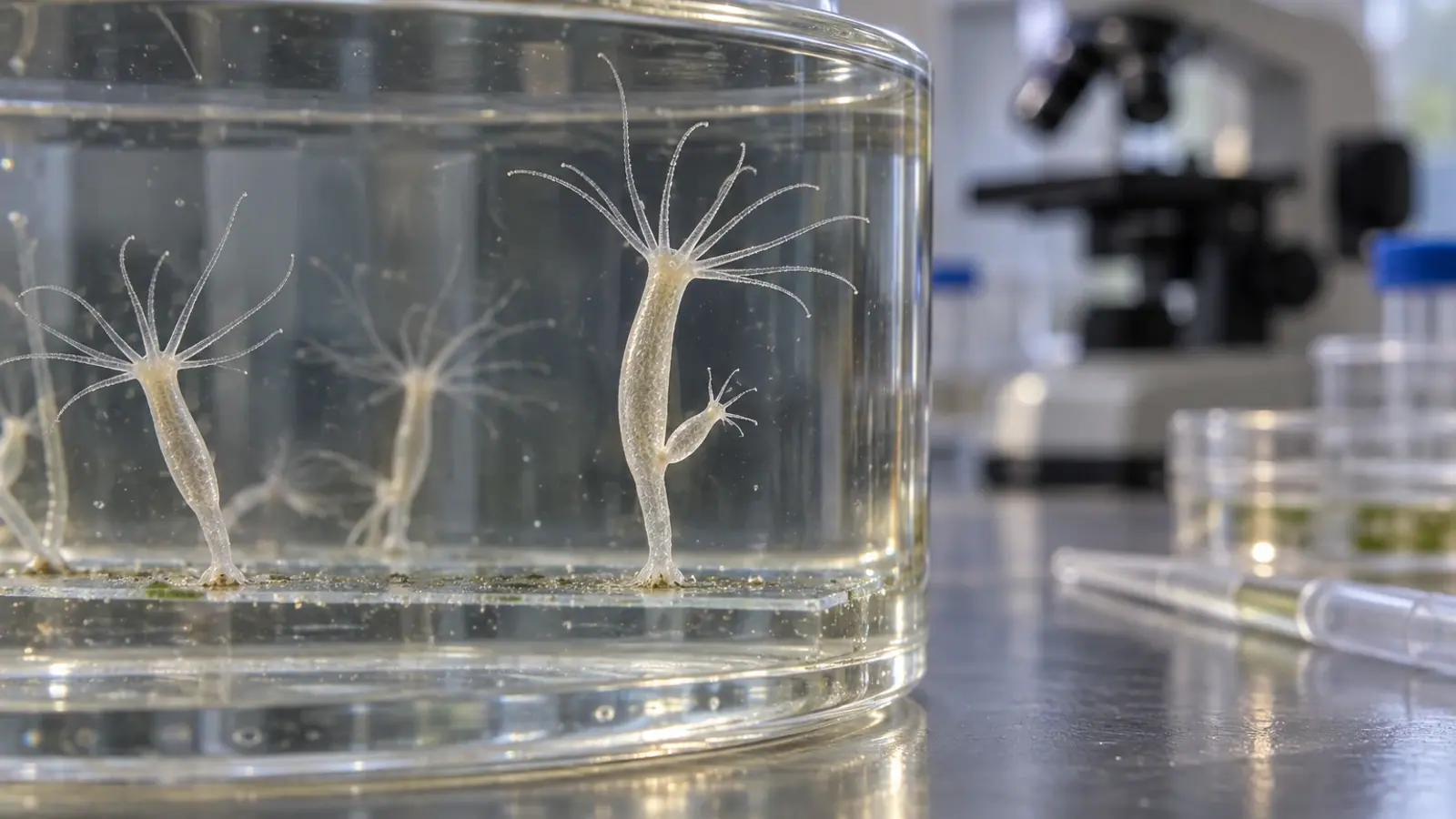
Hydra vulgaris: a model built on constant renewal
This small freshwater cnidarian combines budding, regeneration, stem-cell lineages and whole-body imaging in a model that is simple in shape but not in biology.


- Content type
- Species profile
- Sector
- Research facilities
- Animal group
- Corals and invertebrates
- Keywords
- ReproductionTemperatureStress
A hydra can rebuild a head or foot after bisection, maintain tissues through continuous renewal and reproduce by budding. These features made Hydra a historic regeneration model. Transgenesis, gene knock-down, imaging, genomics and single-cell approaches now connect this capacity with cell states and body-patterning signals.
The model is not interchangeable across laboratories. Several species and strains are used, with differences in genome, reproduction, microbiota and environmental response. Reporting only “Hydra” is insufficient for reproducibility.
Simple anatomy, complex biology
Hydra vulgaris is a freshwater cnidarian with an adhesive foot and a mouth surrounded by tentacles. Two epithelial layers are separated by extracellular matrix. Its nerve net is diffuse rather than centralised, while cnidocytes on the tentacles support prey capture.
The oral–aboral axis continually organises the polyp. After injury, wound closure, positional signalling, tissue remodelling and stem-cell responses rebuild proportionate structures. “Perfect regeneration” should not obscure the sequence of processes or the experimental conditions needed to observe it.
Several stem-cell lineages
Modern descriptions distinguish epidermal and gastrodermal epithelial stem cells, a multipotent interstitial lineage that generates neurons, gland cells and cnidocytes, and a germline lineage. This organisation allows researchers to examine self-renewal, differentiation, migration and homeostasis together.
Renewal capacity does not mean every structure returns through indiscriminate proliferation. Remodelling, cell displacement, cell death and signalling programmes contribute differently according to injury site and time. Conclusions therefore depend on the amputation plane, observation window and markers selected.
Genomes expanded the model
The Hydra genome provided a framework for comparing ancient animal mechanisms, including epithelia, contraction, synapses, pluripotency and axial organisation. Newer resources improve assemblies and resolve expression across cell types.
They do not remove identity problems. Historical names such as Hydra magnipapillata remain in papers and databases. Strain, provenance, resource identifier and nomenclature must travel with the data; otherwise a biological-source difference can be mistaken for treatment effect.
Husbandry shapes phenotype
Temperature, water quality, density, feeding rhythm, prey type and cleaning frequency affect growth and budding. Feeding state is not neutral background: it changes size, physiology and resources available for cellular processes.
Prey cultures require their own records because nutritional quality and microbial load can introduce hidden variation. Associated microbiota must also be distinguished from environmental contamination. Shared water and equipment create routes for cross-contamination between strains.
Budding enables clonal expansion but does not replace identification. Each batch should retain its separation date, parent strain, conditions, interventions and unusual events.
Design an interpretable experiment
A regeneration protocol defines section level, orientation, time zero, temperature, feeding state and outcome criteria. Fixed imaging intervals and magnification reduce ambiguity. Operators should harmonise handling because compression, prolonged exposure and imaging delays can create additional disturbance.
Contemporary controls are essential. Historical series do not control changes in prey, water or strain. Omics studies likewise need batch and physiological-state metadata.
Vetofish can support aquatic-model platforms with culture health qualification, procedures, strain traceability and indicators of welfare and quality. Hydra vulgaris is powerful when its apparent simplicity does not erase the biological variables that organise the experiment.
Persistent contraction, detachment, loss of budding or accumulated debris indicates a drift that requires investigation. Poor prey quality, altered water, excessive density, contamination and unusual handling can produce overlapping appearances. Routine review should combine polyp observation, standardised counts, medium quality and feeding history.
Isolating a suspect batch before sharing pipettes or water protects other cultures and preserves diagnostic options. A physically separate backup reduces the chance of losing an irreplaceable strain. Baselines for attachment, contraction–extension cycles, budding and mortality should be defined for each strain rather than borrowed from another colony.
Exclusion criteria need to be decided before analysis: detached animals, irregular cuts, exceeded imaging delays or non-compliant feeding state. Removing observations after viewing outcomes creates bias. Publishing protocols and metadata helps other groups distinguish biological divergence from methodological divergence.
Staff should document deliberate changes to water, prey and cleaning. Pilot a change on a limited, recoverable part of the colony and retain previous conditions long enough to compare effects. Convenience alone does not show that the new practice preserves health and experimental performance.
Practical training benefits from a strain-specific illustrated reference. It can define the appearance of a normally extended polyp, the accepted density range and the response to detachment or debris. Staff can then compare deviations using the same language before an entire experimental series is affected.
The model also requires proportionate welfare thinking. Avoid unnecessary handling, prolonged exposure outside stable culture conditions and repeated imaging without recovery. Refinement should be assessed through observable outcomes rather than assumed from the animal’s small size or simple body plan.
Repository records should link every published dataset to the exact colony and culture interval. This small administrative step makes later reanalysis possible when taxonomy, genome assemblies or cell annotations are updated.
To move from evidence to action, explore our advice and support service and our expertise for research facilities.


