

Sturgeon: what a positive viral PCR says about skin lesions
Finding viral DNA in a sturgeon does not establish the cause of a skin lesion. A 2023 investigation of juvenile lake sturgeon at a Canadian hatchery shows why PCR, histology and husbandry observations need to be interpreted together.
- Content type
- Practical guide
- Sector
- Aquaculture
- Animal group
- Sturgeons
- Theme
- DiagnosticsDiseases
A conservation hatchery investigation
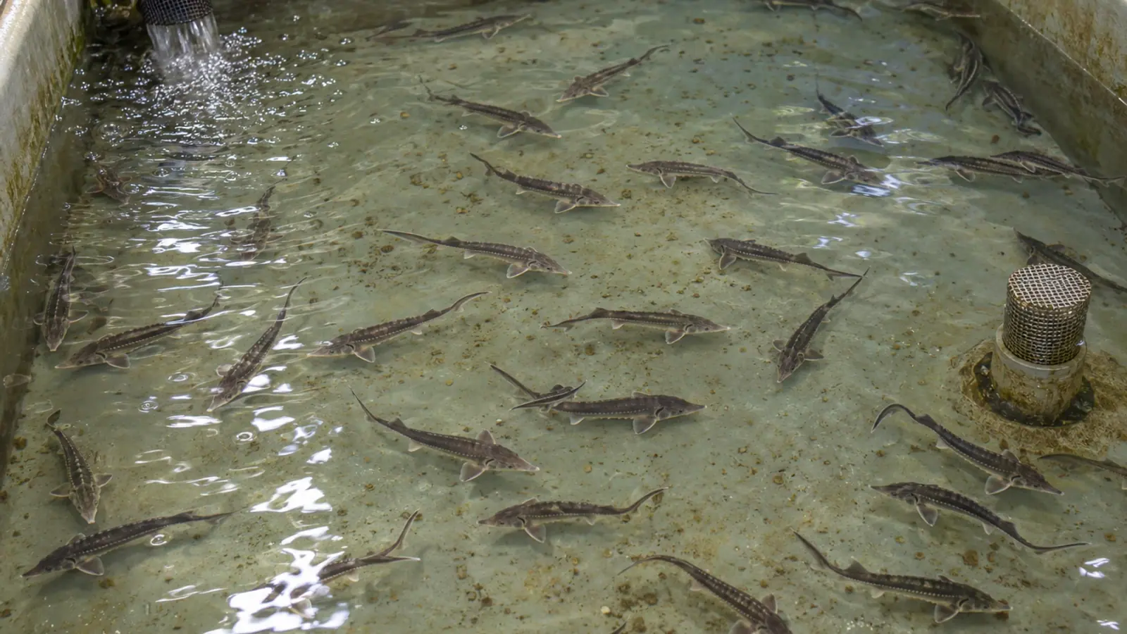
The fish investigated by Clouthier and colleagues were juvenile lake sturgeon, Acipenser fulvescens, reared at Grand Rapids Hatchery in Manitoba, Canada. They came from wild broodstock and were intended for stocking programmes. The facility used recirculating water supplied from wells. This was an investigation in a working hatchery, with families and year classes to follow, rather than an experimental infection study.
Skin abnormalities appeared in the 2017 and 2018 year classes. Fish were approximately 8–11 cm long when lesions were first observed. The growths were pale, smooth and blister-like, occurring on the underside of the abdomen and pectoral fins. They were not deformities of the dorsal scute rows. The authors reported no associated abnormal mortality or behaviour, and the lesions subsequently regressed.
These details define which other cases can reasonably be compared. A different sturgeon species, dorsal ulcers or a sudden rise in deaths would represent a different presentation. A similar photograph cannot establish that the same process is occurring in a new group of fish.
Tissue changes and viral sequences answer different questions
Histopathology showed localised epidermal hyperplasia, with changes including oedema and inflammatory cells. This describes a tissue response. It does not identify its cause on its own. Other organs examined did not show equivalent abnormalities.
Sequencing, followed by PCR and Sanger sequencing checks, identified DNA from an alloherpesvirus designated AciHV-3 in the study. Comparisons of conserved proteins distinguished it from the other sturgeon herpesviruses considered. These results document viral sequences and their genetic relationships. They do not establish that this virus caused all the skin changes observed.
Two findings limit that conclusion. Electron microscopy did not reveal virions in the tissues examined, despite extensive investigation. A suspected cytopathic effect in a sturgeon cell line did not lead to sustained, confirmed virus isolation through passage. The authors explicitly state that further work is needed to establish causality between AciHV-3 and the lesions.
For a hatchery team, the distinction changes how a report should be used. A lesion description and a molecular detection can both be valid while their biological connection remains unresolved. Neither result should be discarded simply because the complete explanation is unavailable.
Why a positive PCR needs clinical context
PCR detects a target nucleic acid in the material tested. It does not directly demonstrate infectious particles, active multiplication or responsibility for a particular lesion. These are separate questions requiring additional evidence.
The study also found related sequences in uninoculated sturgeon cell lines and genomic datasets from several sturgeon species. The authors discuss the possibility of viral elements integrated into the host genome. That hypothesis helps explain why DNA detection requires care. It does not prove inherited integration in every positive fish or establish vertical transmission in the hatchery groups investigated.
Calling the lesions an active, PCR-confirmed viral disease would therefore go beyond the evidence. Treating the finding as mere contamination would also misrepresent the work: viral sequences were identified through several analytical steps. What remains uncertain is how that DNA relates to the fish’s clinical condition.
This is particularly relevant when laboratories and hatcheries exchange short reports. A result described only as “positive” leaves out the tissue sampled, the target tested and the observations that prompted sampling. Those details determine what the finding can support.
Plan sampling around the question
For a skin problem in a hatchery, useful records include species, age, family where known, tank, onset date, lesion location and changes in the number of affected fish. Feeding, behaviour and mortality records connect laboratory findings to the condition of the group. An isolated image rarely provides that context.
Tissue selection and preservation should be agreed with the laboratory before sampling. A piece intended for histology and material intended for molecular testing may require different handling. Fixing all available material before defining the investigation can prevent some analyses. Sending only a molecular sample leaves the tissue architecture undescribed.
Where the veterinarian considers it appropriate, comparing affected and apparently unaffected fish may help place a detection in context. Such a comparison is not proof of causality by itself. Individual identities, groups and sample types must remain traceable so that differences in collection are not mistaken for biological differences.
Temperature and biosecurity without unsupported rules
Cooling preceded some observations, but the pattern was not consistent between year classes. The paper does not demonstrate that a temperature drop triggered the lesions or provide a thermal adjustment that prevents them. Water records help reconstruct a sequence of events; a sequence alone does not establish a mechanism.
Biosecurity decisions should address the actual hatchery situation, including equipment movement, separation of groups, fish transfers and assessment of affected animals. This study validates neither a treatment nor a universal isolation period. It does not establish a single test that can declare a hatchery free from the problem.
The absence of unusual mortality in this investigation should not be applied to every sturgeon skin lesion. The work concerns a particular species and conservation hatchery setting. It is also a 2023 publication, not evidence of a current outbreak in France. AciHV-3 should not be confused with AcIV-E, a sturgeon iridovirus: the similar abbreviations refer to different viral families.
Connect the laboratory finding to the hatchery decision
The contribution of this investigation is the combined documentation of a skin presentation, tissue changes and viral sequences, with causality left open. For an aquaculture team, that approach helps avoid both an overconfident diagnosis and a positive finding with no considered follow-up.
Vetofish supports laboratory testing and diagnosis by relating observed lesions to sampling options and the question to be answered. Defining what each test can establish is the first step towards using its result to decide what happens to the group.


