

Tilapia streptococcosis: confirm the bacterial cause
Signs consistent with streptococcosis do not identify the bacterium responsible. An Indonesian tilapia cage investigation shows why culture, characterisation and molecular findings need to be interpreted together.


- Content type
- Disease profile
- Sector
- Aquaculture
- Animal group
- Fish
- Theme
- DiseasesDiagnostics
- Keywords
- TemperatureStressPCRBiosecurity
Abnormal swimming, declining feed intake and protruding eyes warrant prompt investigation in tilapia. They may accompany systemic bacterial disease, but they do not identify the organism responsible. Establishing a name requires more than matching a familiar clinical picture to a familiar pathogen.
A cage-farm investigation from Lake Sentani, Indonesia, illustrates the problem. The study combined characterisation of bacterial isolates with molecular identification and experimental exposure. Its contribution to a current investigation concerns the methods; it does not estimate present-day prevalence or establish a standard procedure for every farm.
Similar signs led to several bacterial identifications
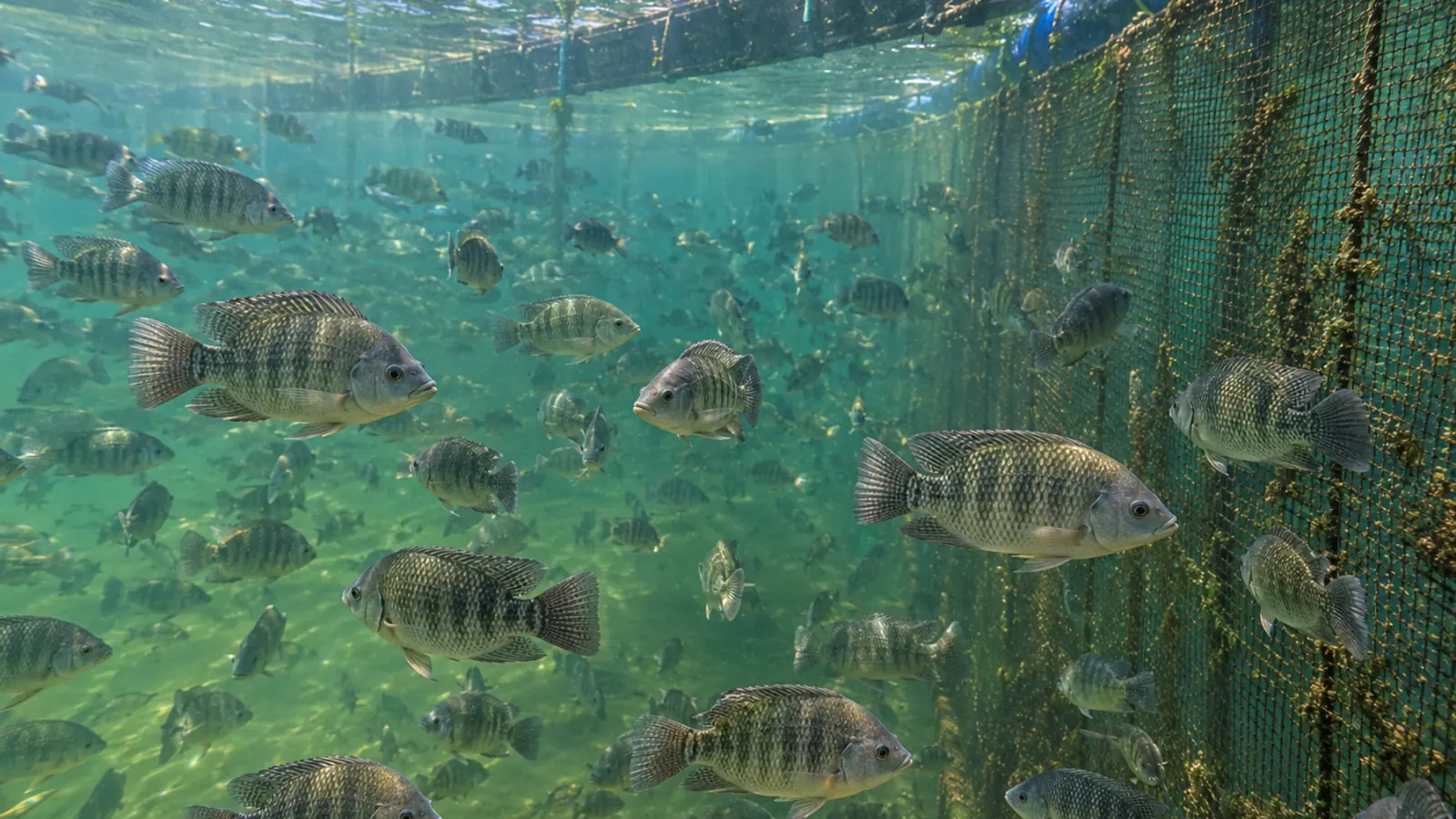
Anshary and colleagues examined 25 clinically affected Nile tilapia, Oreochromis niloticus, from cage culture. Their 2014 paper describes samples from kidney, brain and eyes. Colony appearance and biochemical characteristics suggested related bacteria, without providing a definitive species identification on their own.
The investigators added sequencing of a region of the 16S ribosomal RNA gene and phylogenetic analysis. They assigned four isolates to three species: Streptococcus iniae, Streptococcus agalactiae and Lactococcus garvieae. These are the identifications reported in that study, interpreted using its methods and reference sequences.
Protruding eyes, circular swimming or an opaque cornea could not distinguish those agents clinically. Conversely, isolating several species during an investigation does not establish coinfection in every fish. The authors explicitly stated that it remained unknown whether the pathogens coexisted in the same individual. That uncertainty should survive any practical retelling of the findings.
Experimental pathogenicity is a different question
The researchers also exposed separate tilapia groups to each of the four isolates. Intraperitoneal administration produced disease signs and mortality, with the bacteria recovered from affected fish. This supports their pathogenic potential under the experimental conditions.
It does not measure natural transmission between fish in cages or predict losses on another farm. Injection bypasses normal barriers. Nor did the design compare stocking densities or independently vary temperature to estimate the contribution of those factors.
The practical lesson concerns the diagnostic sequence: characterise the agents, relate the findings to disease and recognise what each method establishes. It does not justify labelling every warm-season mortality as S. iniae infection. A bacterial result may be important while leaving part of the episode unexplained.
Reconstruct the episode alongside the laboratory work
The review by Juárez-Cortés and colleagues describes variation among S. iniae strains, susceptible hosts and interactions with fish defences. It gives biological context for investigating streptococcosis, rather than a universal temperature threshold for every species and production system.
Record temperature, dissolved oxygen and pH with their times and units. A measurement taken after mortality has subsided cannot reconstruct a short equipment failure or an overnight deterioration. Feeding records, daily deaths, grading, transfers, net maintenance and previous treatments help establish the order of events.
Similarly, “stress” becomes useful only when attached to an event that can be examined. Specify what changed, how long it lasted, which fish were affected and what response followed. This keeps environmental and handling factors in the investigation without allowing them to replace an infectious diagnosis.
Match the sample to the diagnostic question
Agree with the veterinary team and laboratory which fish and tissues will be examined and how they will be transported. Freshness and prior antimicrobial exposure can affect culture results. A surface swab or a water sample does not answer the same question as an appropriately collected internal tissue sample.
Culture may provide an isolate for further identification and susceptibility testing. A PCR assay detects a defined molecular target; its interpretation depends on the sample site, lesions, host and analytical controls. A test designed for S. iniae cannot exclude other bacteria. Histopathology can reveal the distribution and character of tissue damage without automatically identifying the bacterial species responsible.
Reports should distinguish an organism being detected, being isolated and being implicated in disease. A positive result may be significant without accounting for every lesion. A negative result from an unsuitable sample may leave the main question unresolved.
Respond to the farm, rather than copying a drug table
Susceptibility results from published isolates are not a prescription for another site. Methods, interpretive criteria and the organisms present can differ. Laboratory susceptibility alone establishes neither clinical effectiveness nor whether a product can be used in the relevant production setting and jurisdiction.
Immediate decisions also concern water conditions and movements that might spread infection. Shared equipment and transfers between groups belong in the biosecurity assessment. Moving compromised fish may itself add a burden, so the proposed action needs to fit the actual installation and the condition of the stock.
Document the working hypotheses, results still pending and the point at which mortality will be reassessed. Preserve findings that do not fit the first explanation. Discordant results may reflect mixed disease, sampling limitations or a separate process that deserves investigation.
Keep the investigation open for a reason
The Lake Sentani investigation shows why similar signs may lead to several bacterial identifications. Combining examination, laboratory methods and a farm chronology helps prevent premature conclusions. The study does not justify assuming coinfection in every fish or attributing mortality automatically to warm water.
Vetofish can help plan diagnostic investigations and interpret the findings in the context of aquaculture operations. The purpose is to identify the next useful examination and proportionate measures for the affected stock, with the uncertainty still visible.


