

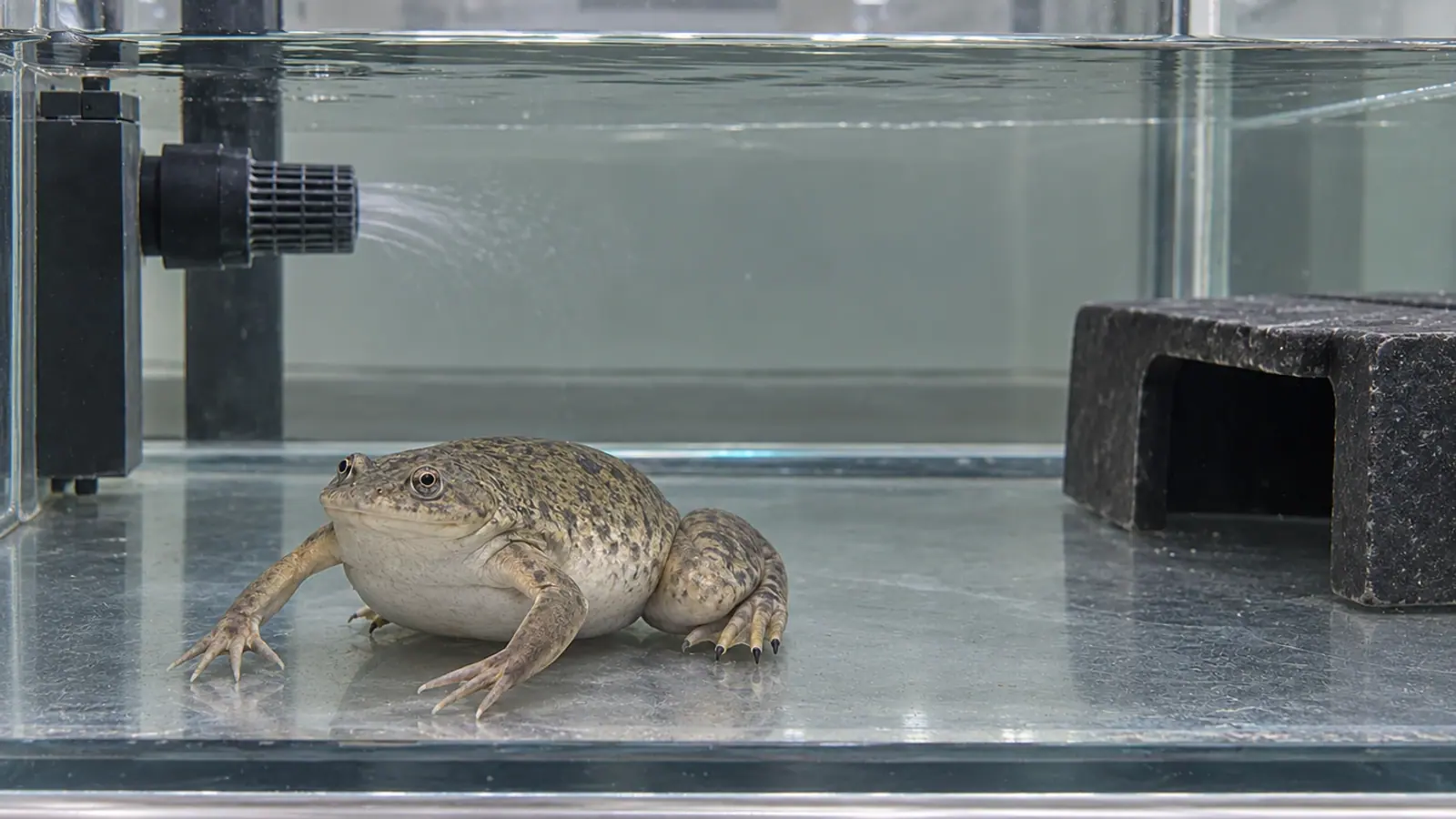
Trichodina in Xenopus: locate it before assigning a cause
A ciliate in a Xenopus laevis urinary bladder does not automatically explain skin lesions. A 2013 case report illustrates why localisation, tissue response and other diagnostic findings need to be assessed together.


- Content type
- Disease profile
- Sector
- Research facilities
- Animal group
- Xenopus
- Theme
- DiseasesDiagnostics
Keep a microscopic finding in its clinical context
Finding an organism in a sick frog provides evidence, but it does not automatically identify the cause of illness. Localisation matters alongside identification. A ciliate in the urinary bladder, a bacterium cultured from blood and a skin lesion answer different diagnostic questions. They cannot be treated as interchangeable evidence of the same process.
Collymore, White and Lieggi’s 2013 case report concerns an adult female Xenopus laevis with skin lesions and reduced appetite. Post-mortem examination found ciliates consistent with Trichodina xenopodus in the bladder lumen, without bladder lesions. Aeromonas hydrophila was cultured from blood. The authors interpreted the ciliate as an incidental finding rather than the explanation for the skin disease and death.
The report remains useful for this specific diagnostic distinction. It does not estimate carriage across laboratory colonies, establish that every bladder ciliate is harmless or provide a treatment trial. Any application to another facility needs to retain those limits rather than converting one case into a general rule.
Identify both the organism and its location
Trichodinids are ciliates with microscopic features, including an adhesive disc and denticulate ring, that can contribute to identification. A name alone does not predict the organism’s effect. Reports should distinguish a compatible identification, a confirmed identification and the extent of the examinations performed.
Histopathology places an organism within a tissue or cavity and describes the response of adjacent tissues. “Present in the lumen” does not mean “invading the wall”. Likewise, absence of a visible reaction in one section does not replace examination of all organs needed for the investigation.
Samples identified by animal and organ help the laboratory retain that context. Images of lesions and a clinical history make findings easier to interpret. The facility should know which tissues were received, what tests were performed and which hypotheses were not assessed. A result is more useful when its limits are visible in the same record.
Reconstruct the sequence before death
Observations made while the animal was alive are part of the diagnostic evidence. Records of appetite, activity, body condition and lesion progression allow laboratory findings to be related to the course of illness. A form completed only after necropsy may miss earlier handling, feeding changes or an accommodation problem.
Inflammation, injury and organism detection need joint interpretation without confusing coexistence with causation. For culture results, the collection site and sampling conditions should remain available. An organism recovered from blood has a different evidential role from one detected in water or on an exposed surface.
Each finding needs to be related to the abnormality it might explain and the available degree of confidence. This preserves alternative diagnoses and helps prevent the investigation ending as soon as the first identifiable organism appears. It also makes disagreement between findings easier to discuss with the laboratory.
Review the group and the aquatic system
An individual record does not replace group monitoring. Check whether other animals show comparable changes, whether several tanks are affected and whether cases follow a shared event. Introductions, breeding, transfers and maintenance interventions should be included in the timeline rather than considered only when the initial tests are inconclusive.
The NC3Rs’ 2024 amphibian care resource emphasises species-specific requirements and the need to validate welfare indicators. Tolerance of variable conditions in the wild does not establish that every fluctuation in a laboratory is acceptable. Monitoring should connect behaviour, feeding and accommodation quality.
A practical checklist can separate water measurements, equipment condition, cleaning and animal observations. It becomes interpretable when methods and observation times are comparable. A single value measured after a correction may not describe the conditions present when the first animal deteriorated. Recording when the correction occurred preserves that distinction.
Select samples to answer the actual question
A negative result can be interpreted only with knowledge of what was sought and which sample was examined. A skin examination does not represent all internal cavities. Conversely, a bladder finding cannot establish the condition of the whole skin surface or contamination across every tank in the facility.
Agree the sampling plan before collection. It may combine clinically affected animals, tissues corresponding to lesions and information about the system. Preservation needs discussion with the laboratory: material intended for culture is not prepared in the same way as tissue intended for histology. Labelling and transport arrangements should be settled while the samples can still be collected correctly.
Conclusions should state their scale: an animal, a group or an installation. Extending an individual finding to the whole colony requires additional evidence. A surveillance programme also needs to explain its detection opportunities and what remains uncertain after a negative check. Such qualification is part of a usable result, rather than an obstacle to action.
Detection alone is not a reason for a bath
The 2013 paper does not establish a validated treatment for bladder infestation. Approaches mentioned for other localisations or trichodinids cannot automatically be transferred to T. xenopodus. This article therefore supplies neither a dose nor a collective bathing instruction.
Treatment decisions need to address the diagnosis and animal condition, with monitoring of response and tolerance. Correcting a husbandry defect may be necessary without proving that it explains the entire illness. Repeated unevaluated interventions can also obscure the sequence and reduce the value of subsequent diagnostic work.
Quarantine and biosecurity need explicit objectives: documenting origin, observing incoming animals, separating relevant flows and retaining equipment traceability. No single check guarantees general freedom from parasites. The measures chosen should match the agents being investigated and the actual system. A plan that cannot be followed consistently provides less information than one whose actions and observations are clearly recorded.
Conclusion: causal attribution needs more than presence
A trichodinid in a laboratory frog needs interpretation through localisation, tissue response and the rest of the diagnostic findings. The published case illustrates that an organism may be present without explaining the principal lesions. Keeping that distinction visible helps direct investigation towards the established problem rather than the most recognisable microscopic finding.
Vetofish support
Our testing and diagnostic service can help define sampling and connect histology, microbiology and husbandry history. For research facilities, the aim is a documented conclusion at the scale actually examined, rather than treating one test as certification of the colony.


