

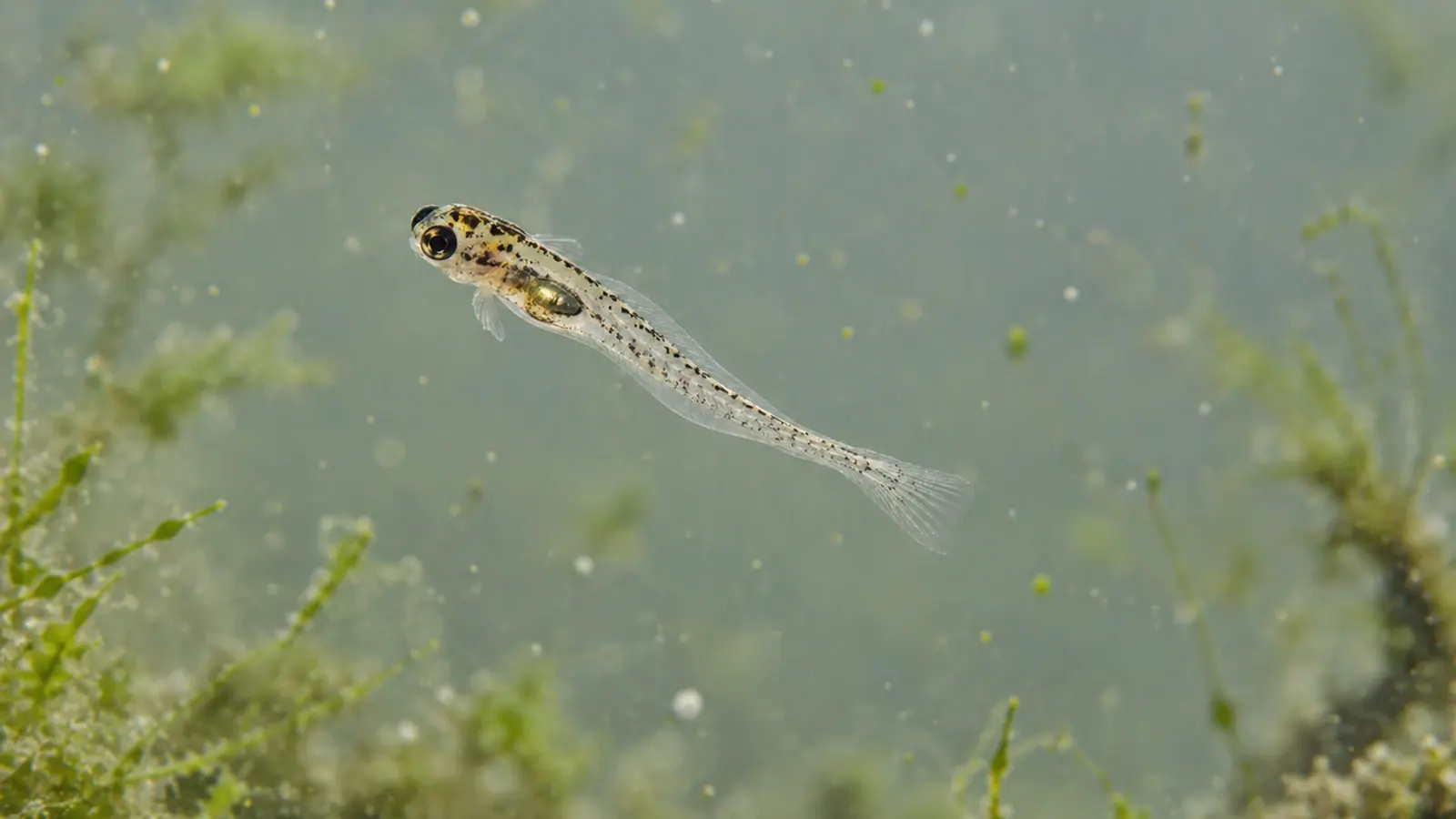
Zebrafish larvae: top-view tracking misses the depth
Two paths that look similar from above can describe different movements through water. A platform published in 2026 combines cameras and mirrors to track zebrafish larvae in three dimensions. It improves the description of movement; interpreting the underlying biology still requires separate evidence.


- Content type
- Scientific news
- Sector
- Research facilities
- Animal group
- Zebrafish
- Keywords
- LightingEthogramAnaesthesia
An overhead camera mainly describes movement in a plane. When a larva rises, dives or tilts its body, part of its movement may disappear from that projection. Two recordings that look similar from above can therefore conceal different positions and movements through the water column.
A study in Communications Biology presents a platform designed to recover that dimension in zebrafish, Danio rerio. Synchronous views provide information about trajectories and selected morphological features. More detailed images describe the movement; establishing its biological explanation requires additional evidence.
Combining overhead and side views
The platform uses twenty-four cameras and a modified plate incorporating mirrors. Its external format follows that of a standard laboratory plate, while some positions are replaced by optical elements. The arrangement records forty-eight compartments, each holding one larva, from two directions.
The top view provides horizontal position. The side view contributes depth and body inclination. Combining them requires spatial and temporal correspondence: an additional image is not enough if it represents another moment or if its geometry is poorly characterised. The reconstruction must relate both views to the same animal and coordinate system.
The researchers therefore assess optical performance, including refraction at the interfaces between air, the plate wall and water. Mirrors also change the light path and focal position. These constraints explain why a three-dimensional tracking platform must be validated as a measuring instrument before its output is treated as a biological response. Greater visual detail is useful only when the source and limitations of the measurement remain clear.
Automated reconstruction is conditional on image quality
The analysis software identifies body landmarks and combines information from the two views to reconstruct position and posture. Its image models are trained on annotated landmarks. Results consequently depend on image quality, annotations and the model’s ability to locate the relevant structures under the conditions being studied.
Automatic tracking is not necessarily complete tracking. In one demonstration, three-dimensional reconstruction succeeded in forty-five of forty-eight compartments. That is a technical performance result for that series, not the animal number for every comparison in the paper. The experimental group comparisons include demonstrations using eight larvae per group.
Excluded frames and recordings belong in the assessment. If failures occur more often in a particular posture or during a certain movement, the retained data may not represent the full behaviour. A high detection rate does not remove the need to examine missing observations or to check that equivalent criteria were applied across groups. Retaining a clear account of those decisions helps distinguish a change in the animal from a change in what the software could measure.
Swim bladder volume is an estimate with visibility limits
The platform also estimates the geometry of the swim bladder, an organ involved in buoyancy. Software segments its appearance in both views, then fits a geometric representation to reconstruct volume. The result is an image-derived estimate rather than a direct observation of every point on the organ’s surface.
When a larva faces towards a camera, its eyes can obscure the structure of interest. Focus also becomes inadequate in some positions. The authors exclude frames that fail their visibility criteria and identify depth of field as a limitation. Those selections need to accompany any comparison of estimated volumes, particularly if groups occupy different parts of a compartment.
A relationship between vertical position and estimated volume does not, by itself, establish the physiological mechanism connecting them. It is also insufficient to diagnose a buoyancy disorder. Developmental stage, animal condition and experimental circumstances provide information that the reconstruction cannot supply on its own. A precise-looking shape should not conceal uncertainty about its biological interpretation.
Retain preparation history and recording rate
The larvae were seven days post-fertilisation. That stage belongs with the result: a method evaluated at this age is not automatically equivalent for adults or other aquatic models. Preparation matters as well. The methods report cold anaesthesia before transfer, followed by recovery, so describing the complete procedure as free from prior anaesthesia would be inaccurate.
Acquisition rate sets another boundary. A short exposure can produce a sharp frame without recording enough separate moments to reconstruct a very rapid movement. The authors note that some brief motor responses are not fully resolved at full-resolution settings. Spatial sharpness and temporal resolution describe different aspects of the recording.
Where tracking output is related to an ethogram, each variable needs a definition: displacement, body inclination, a motor event or occupancy of a zone. Lighting conditions and preparation history should remain attached to the observations. A difference in distance travelled is a locomotor measurement; it does not automatically become a score of anxiety, pain or welfare. The biological interpretation requires evidence relevant to that claim.
Choose the dimension that answers the research question
The main contribution is access to movement outside the overhead camera’s plane. Two-dimensional measurements may remain suitable for some questions, provided their interpretation acknowledges what they omit. Comparing datasets first requires checking that they describe the same dimension of movement rather than assuming that all tracking distances are equivalent.
The paper also demonstrates pharmacological applications. Those findings are not treatment recommendations. Several authors declare links with the company developing the equipment. Those interests, together with the technical limitations, belong in an assessment of the platform; commercial availability does not independently validate every possible use.
For research facilities, Vetofish offers methodological support connecting measurement quality, animal context and interpretation. A useful review states which images were retained, what was excluded, how experimental units were defined and where the conclusion stops. An automated trajectory can then contribute evidence without being asked to carry the entire biological explanation.


