

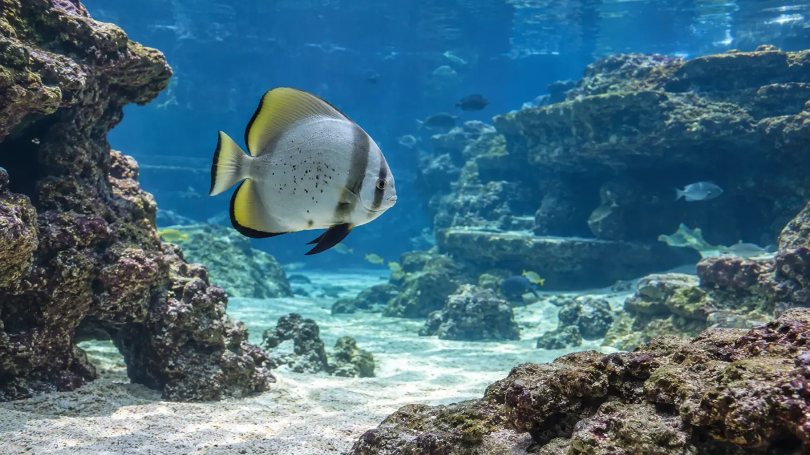
Batfish mycobacteriosis: how far can identification go?
A 2026 report in Platax orbicularis combines granulomas, acid-fast bacilli and sequences related to Mycobacterium marinum. It illustrates how mycobacteriosis can be well supported while the exact bacterial species remains unresolved.
- Content type
- Scientific news
- Sector
- Public aquariums
- Animal group
- Fish
- Theme
- DiagnosticsDiseases
A disease diagnosis can be stronger than a species name
Recognising mycobacteriosis and identifying the exact bacterium are different diagnostic achievements. In May 2026, Kim and colleagues reported an orbiculate batfish, Platax orbicularis, found dead in a South Korean public aquarium. The fish measured 33 cm and weighed 1.3 kg. Its examination produced converging evidence of systemic disease, while leaving an important limitation on the organism’s precise identity.
The report adds an observation in this host and an example of an investigation performed on tissues that had already been fixed. It is neither a survey of mycobacterial frequency in public aquariums nor a treatment trial. For aquarium teams, the case shows how sample preservation shapes both the available tests and the precision of their conclusions.
Establish the relationship between bacteria and lesions
At necropsy, the gills contained pale to whitish nodules, some merging together, with damage to the normal arrangement of the filaments. Histopathology subsequently demonstrated granulomas in the gills, spleen and kidney. The spleen and kidney showed the most severe changes, including areas of necrosis surrounded by inflammatory cells.
Ziehl–Neelsen staining revealed abundant acid-fast bacilli within the lesions. Their location matters because it connects the bacterial finding with tissue damage. Detection in aquarium water alone would not carry the same diagnostic meaning. Granulomas together with intralesional acid-fast bacilli strongly support a mycobacterial explanation, but microscopy cannot by itself establish the exact bacterial species.
Understand what fixed tissue preserves
The available samples had been fixed in formalin and embedded in paraffin, commonly abbreviated to FFPE. This preserves organ structure for histological sections. The same processing can fragment DNA and introduce cross-links that make extraction and amplification more difficult. A tissue archive can therefore remain informative while imposing limits on the molecular work that can be performed.
The researchers nevertheless amplified fragments of the 16S ribosomal and hsp65 genes using nested PCR, then sequenced the products. They included a no-template control and separated preparation, amplification and post-PCR handling. These precautions describe the analytical arrangement used for the case. They do not remove the constraints imposed by short sequences and the absence of alternative fresh material.
Sequence similarity is not a complete identification
The recovered fragments were most closely related to reference sequences of Mycobacterium marinum. Similarity for hsp65 reached 99.5% in a gill sample and 100% in a kidney sample. Those figures describe the fragments examined, rather than the whole bacterial genome. They cannot be read as definitive evidence that every relevant part of the organism is identical to a named reference species.
The phylogenetic analysis used the highest-quality kidney-derived hsp65 sequence. Other, shorter fragments were excluded from that tree, and branch-support values were not displayed. The authors consequently use the cautious designation “M. marinum-like organism”. This wording preserves the useful result while making clear that the investigation did not achieve a definitive species-level identification.
Missing culture leaves a diagnostic gap
No fresh tissue suitable for mycobacterial culture had been retained. The investigation therefore had no living bacterial isolate for complementary confirmation or further characterisation. Molecular results from FFPE material cannot replace that option. Instead, the case shows how archived samples can contribute when the initial preservation choices have already restricted what is possible.
For a new case, the veterinarian and laboratory can plan separate samples for the intended examinations. Material for histology and material for microbiology may require different handling. The organs, containers and transport arrangements need to be agreed before dispatch. Placing every available tissue in the same fixative can close a useful diagnostic route before the receiving laboratory has assessed the question.
Avoid inventing the aquarium’s exposure history
Detailed records of water quality, stocking density, filtration, quarantine and other fish sharing the system were unavailable. The report cannot establish how this batfish encountered the organism, how long it had been present or whether other animals were affected. One necropsy provides no basis for a judgement about the aquarium’s overall husbandry standards.
Granulomatous inflammation also requires a differential diagnosis. Other bacteria, some fungal agents, parasites and foreign material can produce lesions of this broad type. In the reported fish, intralesional bacilli and molecular findings reinforce the mycobacterial interpretation. The diagnostic strength comes from their agreement with the tissue changes, rather than from the gross appearance of a nodule alone.
Keep occupational implications proportionate
M. marinum is a recognised zoonotic pathogen, but this case contains neither an assessment of human exposure nor a reported infection in an aquarium employee. The missing isolate and incomplete species identification make that distinction particularly relevant. The investigation does not quantify any individual worker’s risk or demonstrate transmission from this fish to a person.
A suspicion can still justify reviewing tasks involving water and tissues with those responsible for occupational prevention and biosecurity. The laboratory should receive the relevant diagnostic concern. This supports appropriate planning of sample handling, while avoiding an unobserved human transmission narrative based solely on a sequence related to M. marinum.
Conclusion and Vetofish support
The batfish case supports a precise conclusion: systemic mycobacteriosis associated with an organism closely related to M. marinum, with limits on definitive species identification. The value lies in the combined evidence from lesions, staining and molecular analysis, and in an honest account of the missing tests.
Vetofish can help plan sample selection, preservation and interpretation through its analytical and diagnostic service for public aquariums. Preparing the clinical record and sampling plan early helps preserve options before tissue is fixed or exhausted.


