

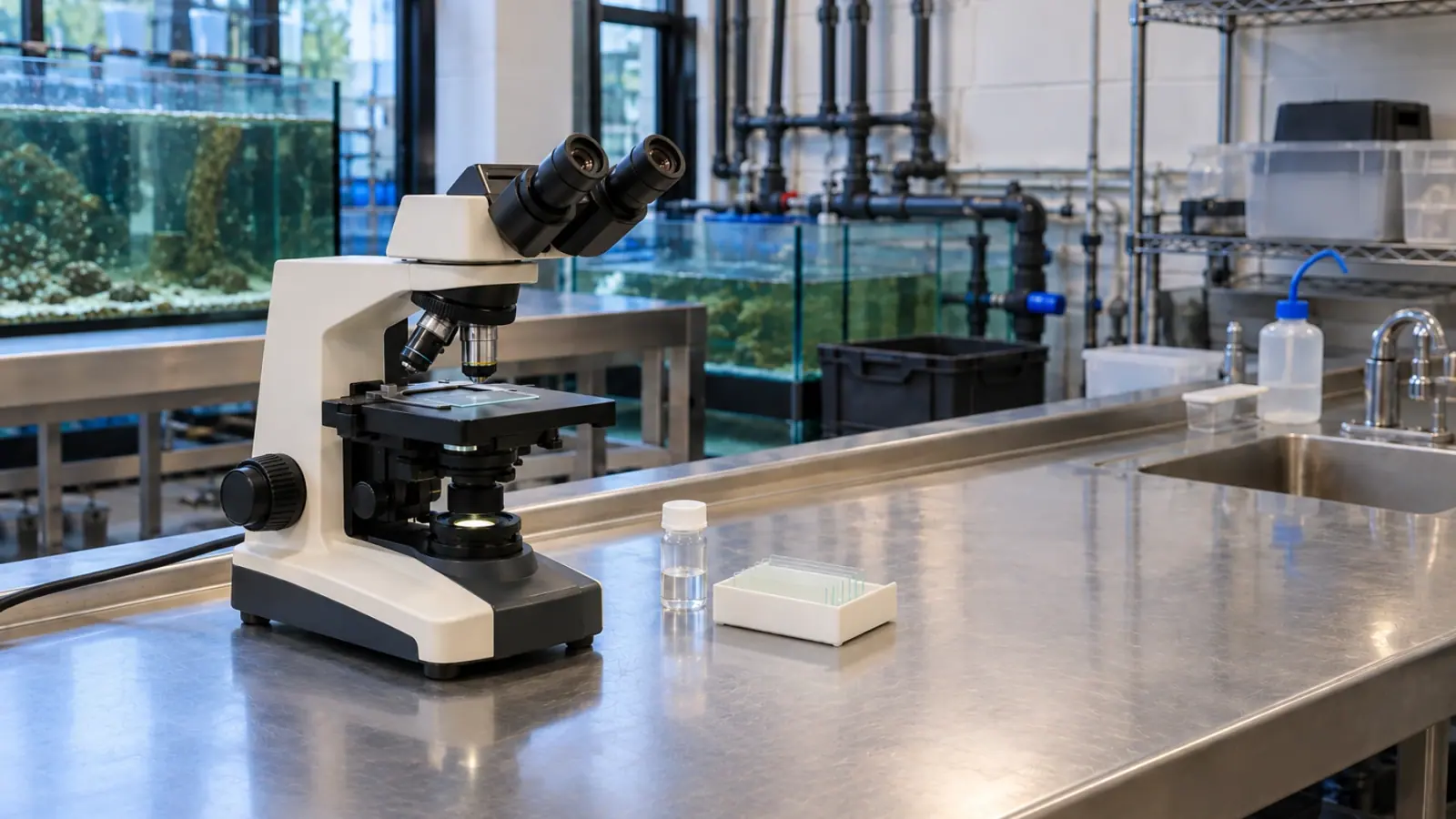
Fish skin and gills: what a wet mount can reveal
Fresh skin and gill preparations can provide an early diagnostic lead in aquarium fish. Sampling site, preparation quality and clinical context determine what the result means and which further tests are needed.
- Content type
- Practical guide
- Sector
- Public aquariums
- Animal group
- Fish
- Theme
- DiagnosticsDiseases
- Keywords
- HistopathologyPCRBiosecurity
Start with the diagnostic question
Rubbing, altered breathing or a change in skin appearance does not identify a particular parasite. Water conditions, infection and interacting problems can produce similar clinical signs. In a public aquarium, microscopy is useful because it lets the team compare an observation with material from a selected site rather than assign a cause from appearance alone.
A wet mount examines fresh biological material without the complete fixation and processing used for tissue sections. Depending on the sample, it may reveal organisms, mucus and tissue structures. Keeping material close to its initial condition offers useful information, but preparation and interpretation still require appropriate skills.
UF/IFAS describes a diagnostic investigation that combines system history, water measurements, examination of the fish and further laboratory tests. A fresh preparation belongs within that investigation. It does not remove the need to check oxygen, temperature or the events preceding the first clinical signs.
Choose a site before arranging a capture
Skin and gills are different sampling sites. Material taken from the body surface does not automatically describe conditions in respiratory tissues. The veterinarian selects a method in relation to species, size, clinical condition and the question that needs answering. The least disruptive useful examination may differ between animals in the same collection.
Useful documentation begins before the slide is made. It connects the sample with a tank, an animal, a precise site, the observed signs and any previous treatment. Where several sites are examined, keeping preparations separate preserves the origin of each finding. This becomes particularly valuable when images or results are later discussed with a diagnostic laboratory.
Sampling also needs a welfare plan. Obtaining material is not a justification for prolonged restraint, dry skin or repeated captures without a defined purpose. Preparing equipment and arranging the reading beforehand can reduce unnecessary handling. The practical method, including any sedation or other support required, belongs in the veterinary procedure rather than a general aquarium instruction.
Describe what is visible before assigning a name
The UF/IFAS account of freshwater white spot disease provides a useful example. White spots alone do not confirm Ichthyophthirius multifiliis, and some microscopic stages can resemble other ciliates. Identification depends on the features actually visible in the preparation and on the case context, rather than on a rounded shape or movement alone.
An organism should therefore be recorded with its site, appearance and the limits of identification. A photograph or short video can assist consultation when scale and sample origin are retained. An unclear image cannot establish a distinguishing feature that was not visible in the original examination. Uncertainty should remain in the record until it can be resolved.
Abundance needs equally careful wording. A parasite seen on one preparation has been demonstrated in that sample; it has not thereby been shown to be abundant throughout the tank population. The number of animals examined, the sites sampled and whether the finding recurred should accompany the interpretation.
Consistent terms help the team avoid accidental changes in meaning. “Organism observed”, “identification suspected” and “cause established” describe different levels of conclusion. The report should use the level justified by the examination. A preliminary finding can be useful for planning the next test without being presented as a complete explanation of the clinical problem.
Decide what further tests could establish
Histopathology examines tissue architecture after processing, whereas a wet mount provides a different kind of view. Culture, molecular tests or additional examinations may be needed for particular hypotheses. None of these methods answers every possible cause of skin or gill disease, and the choice should follow the question rather than habit.
A negative result first means that the target was not identified under the conditions of that examination. It does not demonstrate that the collection is free of disease. Sampling site, sample condition, previous treatment and reading quality may restrict what can be concluded. Persistent signs with an uninformative preparation call for reassessment of the diagnostic approach.
A positive finding may also leave part of the investigation unresolved. More than one factor can contribute to clinical signs, and water conditions remain relevant after an organism has been detected. Diagnosis connects findings with plausible explanations; it should not stop automatically at the first visible structure under the microscope.
Arrange the next step while samples are still useful
Before collecting all material in the same way, the team should discuss possible further tests with the laboratory. Preservation suitable for one analysis may prevent another. Fixing every sample, for example, removes options that require living material. Planning separate sample pathways is easier before collection than after the material has been processed.
A concise report can distinguish the observation, its interpretation and the proposed next check. Another clinician should be able to recover which fish and site were examined, the method used and the remaining uncertainty. Overconfident wording can prematurely close an investigation that still has unresolved environmental or clinical questions.
Where fish are deteriorating, welfare and correction of identified hazards remain priorities. Preserving diagnostic value does not mean postponing essential care. Veterinary and aquarium staff need to coordinate those needs and record the timing of interventions, so that later results can be interpreted in light of what happened.
Keep the early answer useful
A wet mount is valuable when it informs an action: investigate a suspicion, arrange a complementary test or reconsider an explanation. Speed does not replace competent interpretation or subsequent follow-up. The team still needs to judge whether the progress of the fish fits the working diagnosis.
For public aquarium teams, our testing and diagnosis service can help frame sampling questions, coordinate examinations and interpret results alongside tank history. Any treatment decision follows that assessment rather than a universal recipe or a photograph considered in isolation.


