

A cloudy fish eye: locate the abnormality first
Corneal clouding, a cataract and a protruding eye describe different abnormalities. In public aquariums, locating the change and assessing feeding help direct the examination. A small surgical case series shows why outcome estimates need careful interpretation.
- Content type
- Practical guide
- Sector
- Public aquariums
- Animal group
- Fish
- Theme
- DiagnosticsDiseases
A white-looking fish eye is an observation that still needs anatomical localisation. The cornea is the transparent surface in contact with the water; the lens lies inside the globe. Clouding may involve either structure. A protruding eye describes a change in position or size, rather than establishing the cause of the abnormality.
Public aquarium teams can use this distinction to organise observations and prepare a veterinary examination. It also keeps the appearance of the eye separate from its functional consequences. A conspicuous change and a fish’s ability to find food do not necessarily deteriorate at the same rate.
Start with a description that can be checked
The initial record should identify the animal, the affected side, the date and the observed progression. An overall photograph and a useful view of the eye provide complementary information. Reflections and viewing angle can change the impression of clouding, so a single image cannot replace an examination.
Exophthalmos means abnormal protrusion of the globe. It is not a diagnosis of bacterial infection. A unilateral change, bilateral involvement, a witnessed injury and several affected fish suggest different lines of investigation. These observations should accompany the referral without being converted into an assumed cause.
Recent handling, exhibit incidents and changes in general condition also belong in the record, alongside available water measurements. Their purpose is to establish context. A temporal association with a husbandry change may guide investigation but does not prove that the change caused the eye problem.
Locate the opacity before calling it a cataract
A cataract is an opacity of the lens. It needs to be distinguished from corneal oedema, a surface lesion and deeper ocular abnormalities. More than one structure may be affected, so anatomical localisation does not necessarily identify a single disease process.
The 2026 INHAND chapter on fish special senses provides terminology for ocular lesions, separating changes in the cornea, lens, retina and other structures. It also draws a useful distinction between clinical and microscopic language. Cataract is a clinical diagnostic term; a histopathology report should describe the tissue lesion observed, such as degeneration of lens fibres.
Shared terminology helps clinicians and pathologists investigate the same anatomical question. The reference is not a vision test or a treatment protocol. Microscopic findings can clarify tissue changes without reconstructing precisely what the fish could see before examination.
Assess the consequences for the fish
Food intake, access to food, body condition and repeated injuries help establish functional impact. A ration disappearing from a group exhibit does not demonstrate that the affected individual has eaten. Targeted observation is needed to distinguish access to food from the performance of the group as a whole.
Position in the water column, collisions and interactions with other fish can add information. None provides an isolated measurement of visual acuity. Loss of appetite or unusual swimming may also arise from a general health problem and should remain part of the wider clinical assessment.
Intervention decisions depend on the lesion, its consequences and the animal’s condition. Appearance alone does not predict outcome. A relatively subtle change may still warrant prompt investigation when feeding or physical integrity is deteriorating.
What a small surgical series can tell us
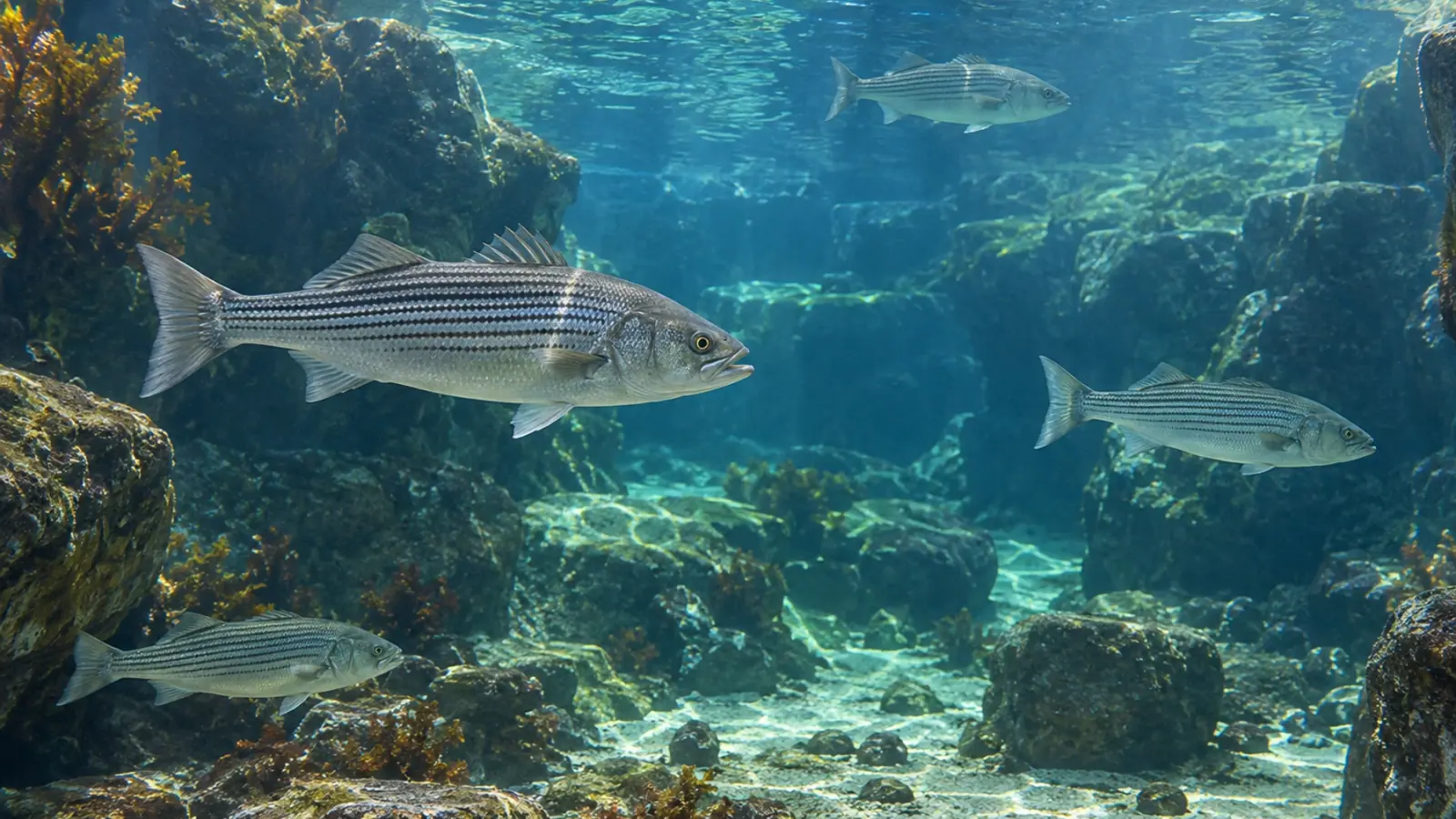
Pujol and colleagues described eleven adult fish at the Aquarium du Québec examined between 2015 and 2021. The series included striped bass, Morone saxatilis, Atlantic wolffish, Anarhichas lupus, and spotted wolffish, Anarhichas minor, with mature cataracts. Four fish underwent bilateral surgery, giving a total of fifteen operated eyes.
After lens extraction, eight of the eleven fish resumed feeding and recovered good body condition. Three retained visual impairment associated with anorexia and were euthanised. Postoperative corneal oedema was common and resolved over the follow-up described by the authors.
This was a retrospective series without an untreated comparison group. Eight feeding recoveries out of eleven fish cannot be applied as a universal probability of success. Fifteen eyes are not fifteen independent animals, and a favourable clinical course does not demonstrate identical restoration of vision in every individual.
The findings support an informed discussion about selected cataract cases. They do not establish the prognosis of another species or an animal with a different underlying problem. Feeding, general condition and ocular findings need to remain separate outcomes when discussing expectations with the team.
Match each examination to its purpose
In the case series, clinicians combined general physical and ophthalmic examinations, with ocular ultrasound where needed. Ultrasound can document some internal structures when opacity restricts direct observation. Performing it does not, on its own, identify the cause or replace all other examinations.
Any examination requiring capture or anaesthesia should be planned for the species and the animal’s clinical state, with appropriate monitoring. Preparation helps obtain useful information while avoiding unnecessary repeat handling. The case series should not be used as an unsupervised surgical or medication recipe, and no such instructions are provided here.
The clinical record should retain photographs, the anatomical location, working hypotheses, examinations and subsequent feeding observations. Consistent descriptions make follow-up more informative: a change in wording between shifts should not be mistaken for a change in the fish.
Conclusion and Vetofish support
A cloudy eye calls for anatomical localisation, assessment of functional consequences and investigation of possible causes. A small surgical series can inform that discussion without determining an individual outcome.
Vetofish’s aquatic health expertise can support public aquarium teams in preparing observations and investigations. Bringing the ocular findings together with general condition and individual food intake helps turn an imprecise report of a white eye into a more useful clinical question.


