

Fish ultrasound: what lies behind a fluid-filled lesion?
A fluid-filled appearance cannot establish the cause of a lesion or exclude a tumour. A common carp case from a public aquarium illustrates how follow-up imaging, complementary modalities and careful sampling can change the diagnostic interpretation.
- Content type
- Practical guide
- Sector
- Public aquariums
A fluid-filled structure on a fish ultrasound is a useful description, but it is not yet an explanation. It can help locate an abnormality and select the next examination. It cannot, on its own, distinguish a cyst from an inflammatory collection or the fluid component of a different lesion. Even an aspirate containing no identifiable tumour cells may leave that distinction unresolved.
A common carp case reported by Hyatt and colleagues in 2013 demonstrates this problem in a public aquarium. The sequence of findings shows how the interpretation changed. One historical case cannot establish disease frequency or estimate the diagnostic accuracy of ultrasound across aquarium collections. It can, however, help teams recognise what an individual result does and does not answer.
Describe the anatomy before naming the lesion
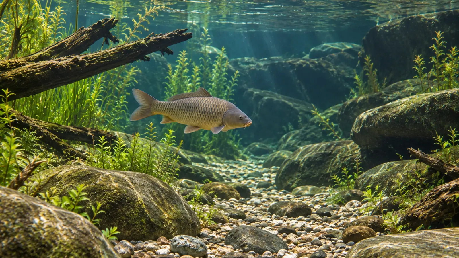
The male common carp, Cyprinus carpio, weighed 7.5 kg and measured 66 cm. It was housed at Georgia Aquarium and presented with a focal right-sided swelling and erratic swimming. Initial ultrasound showed an anechoic, apparently fluid-filled structure near the body wall. The differential diagnoses included seroma, trauma, abscess, cyst and neoplasia.
Its apparent location did not establish its origin. A useful report distinguishes the coelomic cavity, body wall and adjacent structures, then records which relationships are visible and which remain uncertain. Anechoic means that little echo is returned under the examination conditions. It does not describe the microscopic identity of the tissue, and a dark region on an ultrasound image is not evidence of a benign process.
Record the dimensions, shape, contents, wall and identifiable anatomical relationships. Areas that cannot be adequately assessed belong in the report as well. Probe selection and the available examination window depend on the fish and the question. The equipment used in one large carp cannot be treated as a universal imaging protocol for smaller teleosts or other aquarium species.
A fluid sample may not represent the underlying tissue
The clinicians removed 290 mL of serosanguineous fluid. Cytology mainly revealed degenerate red cells against a proteinaceous background, without identifiable neoplastic cells. Aerobic and anaerobic cultures were negative. A seroma was initially adopted as a working diagnosis, but the fluid accumulated again.
Those findings describe a particular sample examined by particular methods. A negative culture does not establish absolute sterility, and failure to find tumour cells does not exclude a tumour. Fluid and the tissue surrounding or producing it may provide different information. The question is therefore whether the material obtained was capable of answering the clinical question, rather than whether a result can be labelled reassuring.
An ultrasound-guided aspirate of the solid component later in the investigation was also non-diagnostic. Targeting an abnormal region does not guarantee that a sample contains the features needed for classification. The next decision should consider sample quality, progression of the lesion and the cost of another invasive procedure. Repeatedly obtaining little information is a reason to review the plan.
Follow-up can reveal a different problem
At the examination one week after presentation, ultrasound showed a more distinct capsule and a small solid component close to the vertebral column. Subsequent examinations documented enlargement of both the cavity and the solid component, with changes in its appearance. The initial impression of a predominantly fluid-filled abnormality therefore became only part of the explanation.
A repeat examination needs an explicit purpose. Comparable landmarks, orientation and archived measurements help determine whether a structure has grown, whether its wall has changed or whether previously obscured tissue can now be evaluated. Images without reliable reference points may give an uncertain impression of progression, even when each individual image is technically readable.
The interval should follow the clinical condition and the decision that another scan could change. Repeated capture and, where needed, anaesthesia impose their own burdens. Recovery and welfare monitoring belong in the assessment of each session. A fixed schedule is not a substitute for deciding whether the information expected from another examination justifies the handling.
Combine modalities while keeping their roles distinct
Radiographs in this case showed lysis and misalignment of neighbouring vertebrae. Bacterial osteitis and neoplasia were among the interpretations considered. Progression despite management strengthened concern about neoplasia. The fish was eventually euthanised. The MRI and computed tomography described in the paper were performed after death, a distinction that matters when presenting the diagnostic sequence.
Necropsy and histopathology established a locally aggressive haemangiosarcoma. Imaging characterised extent and anatomical relationships; tissue examination classified the lesion. This was not an immediate imaging diagnosis and does not show that a single modality outperforms all others. The complete record is useful precisely because the earlier results were incomplete.
An eventual biopsy in another fish should be selected for its likely contribution and clinical value. It is not an automatic requirement for every fluid-filled structure. Surveillance, sampling or a different imaging modality may be appropriate, depending on risk, available expertise and the action that would follow the result. An examination plan should make that intended action explicit.
Keep the living animal in the assessment
Stephen Smith’s review of fish welfare in public aquariums recommends combining behavioural, physiological and performance indicators. Feeding, swimming, social interactions and body condition therefore provide context for an imaging record. Stability in a measured structure does not establish good welfare, while a striking image alone does not determine an individual fish’s prognosis.
The practical conclusion is to retain a reasoned differential diagnosis and a purpose for each examination. Vetofish can help organise the imaging record, discuss sampling options and interpret findings with the care team through our diagnostic testing service, as part of our support for public aquariums.


