

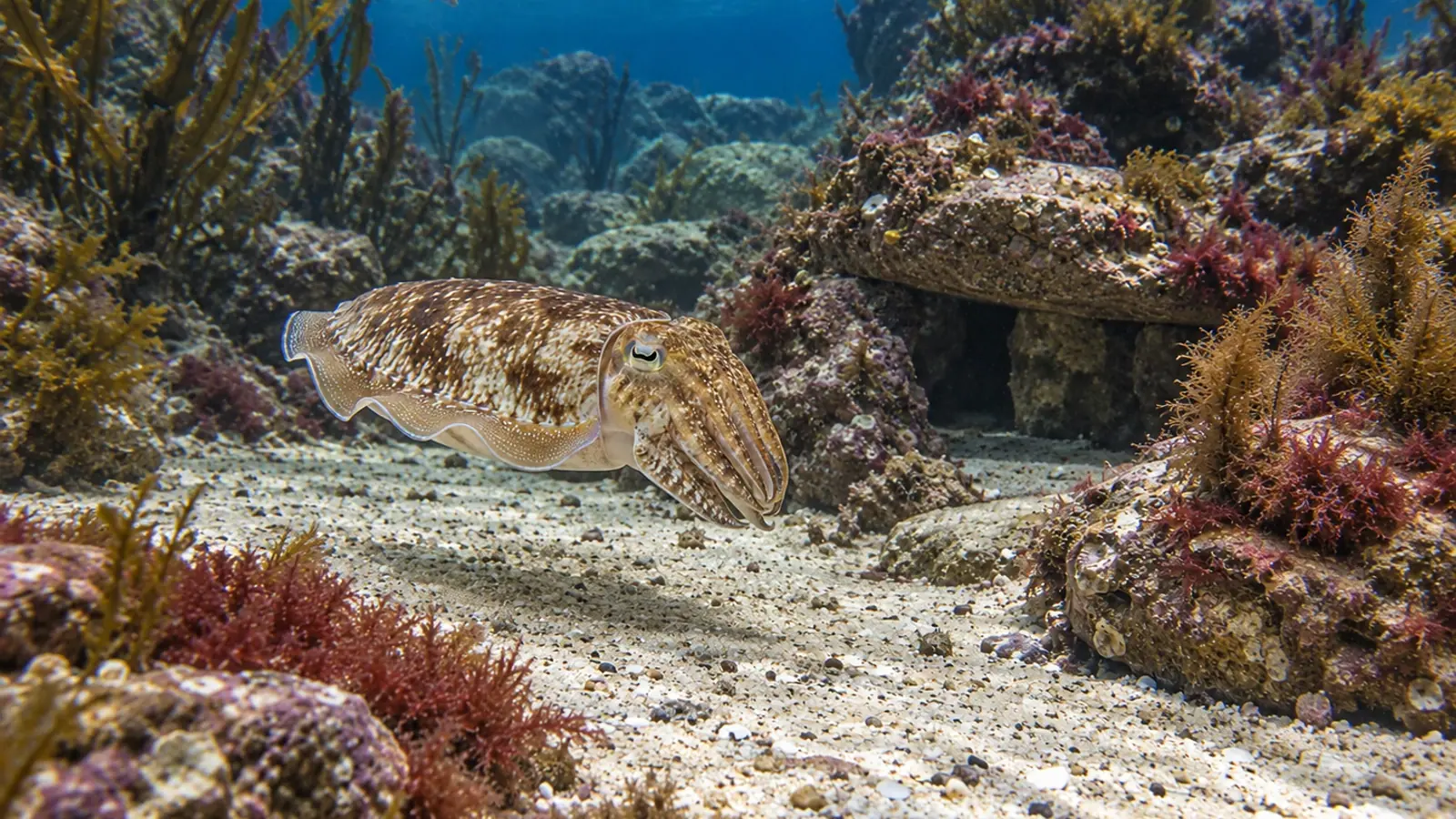
Cephalopods: eye swelling can involve the white body
An apparently swollen eye does not identify the affected tissue. A retrospective series in common cuttlefish and hummingbird bobtail squid highlights changes in the white body, an immune-cell-producing organ around the eye. Aquarium teams can use this finding to broaden assessment while keeping the cause of each case open.
- Content type
- Scientific news
- Sector
- Public aquariums
- Animal group
- Cephalopods
- Keywords
- HistopathologyStressTemperature
Assessing an apparently prominent eye in a cephalopod means considering more than the eye surface. Changes in prey capture, mantle condition or the two sides of the head may provide complementary information. External appearance can establish that something has changed without identifying the affected tissue or the cause.
A study published in the Journal of Aquatic Animal Health in June 2026 links clinical observations with microscopic lesions in two species. It describes changes in the white body, a structure around the eye.
Read the case series as a selected group
Van Eijk and colleagues retrospectively examined submissions from a research facility to a pathology service between 2017 and 2024. Their series comprised 19 common cuttlefish, Sepia officinalis, and six hummingbird bobtail squid, Euprymna berryi. Cases were included because an ocular or periocular abnormality had been recorded clinically or identified in the tissues.
The 25 animals provided 50 eyes for examination, together with clinical records and specimens suitable for gross and histopathological assessment. Selection matters when interpreting the proportions. They describe submitted cases with abnormalities, rather than the frequency of eye disease among all animals at the facility or across public-aquarium collections.
Applying the findings to an aquarium therefore requires a case-based comparison. Species, age, housing and the circumstances in which a sign developed remain relevant. The original animals were laboratory-housed, and the study was not a survey of aquarium populations.
Why the white body belongs in the assessment
The white body is associated with the eye region and contributes to the production of haemocytes, the defence cells of cephalopods. Its location helps explain why a change in its size may matter when an eye appears displaced, even if the eye tissues themselves have no major lesion.
Bilateral white-body hyperplasia was recorded in 15 animals in the series. Hyperplasia means an increase in the number of cells within a tissue. The authors interpreted this finding as potentially reactive to inflammation elsewhere in the body. Skin or gill lesions could accompany changes around the eyes.
That is an interpretation of associated lesions, not an established explanation for every case of apparent eye protrusion. An external photograph cannot confirm white-body hyperplasia. It can document asymmetry, progression or a change in appearance that warrants a broader clinical examination.
The distinction is useful during a discussion between keepers and the veterinarian. A description of a prominent left eye, for example, preserves what was observed. Naming a particular underlying lesion requires further evidence.
Several tissues can produce a visible change
The study also describes inflammation within the eye, cataracts and less frequent abnormalities. A whitish appearance is consequently not a diagnosis in itself. The visible opacity, its suspected location and the lesion eventually identified should remain distinct in the record.
Histopathology helps connect appearance with tissue organisation. In this study it was performed on animals that had died or been euthanised during their clinical management. It is not presented as a routine live-animal check for every minor visual change.
The results did not establish one infectious agent shared by all affected eyes. Bacteria recovered from some tissue samples do not demonstrate infection in every eye with a lesion. Associations involving parental lineages likewise require further investigation before a hereditary cause can be assigned.
For an aquarium team, these distinctions prevent an immediate jump from a sign to an assumed treatment. The examination plan depends on the species, general condition, progression and the information that can be obtained without making the animal’s situation worse.
Follow the animal without adding unnecessary handling
Initial monitoring can use observations made under comparable conditions: which side is affected, the apparent position of the eye, opacity, orientation towards prey and successful feeding. Missing a prey item can have several explanations and does not, on its own, establish blindness.
Dated photographs taken from similar angles and with comparable lighting help assess change. Reflections from the viewing window and changes in posture need to be recognised so that they do not create an impression of swelling. Earlier photographs of the same individual may be more informative than a single comparison between different animals.
The wider record should include mantle and arm condition, ventilation and usual activity. Staff can also place temperature records and husbandry events alongside the clinical timeline. In the study, limited variation in environmental conditions and incomplete data constrained the identification of risk factors. Failure to demonstrate an association in that dataset does not exclude an environmental contribution in a new case.
Monitoring itself has a welfare cost when it requires disturbance. Repeated capture to obtain a clearer photograph may add stress without resolving the clinical question. The veterinarian and keepers can agree which observations matter most, when to reassess and which changes require a faster response.
Broaden the examination while keeping the cause open
The series adds the white body to the clinical reasoning around an apparently protruding eye in these cephalopods. Its contribution is anatomical and descriptive: a visible sign can reflect neighbouring tissues or a more general condition. It does not supply a remote diagnosis, a collection-wide prevalence estimate or a universal treatment.
Vetofish works with public aquariums through diagnostic assessment and laboratory support, connecting keeper observations with the animal’s examination and appropriate investigations. A shared photographic record and timeline help define the question before choosing how to answer it.


