

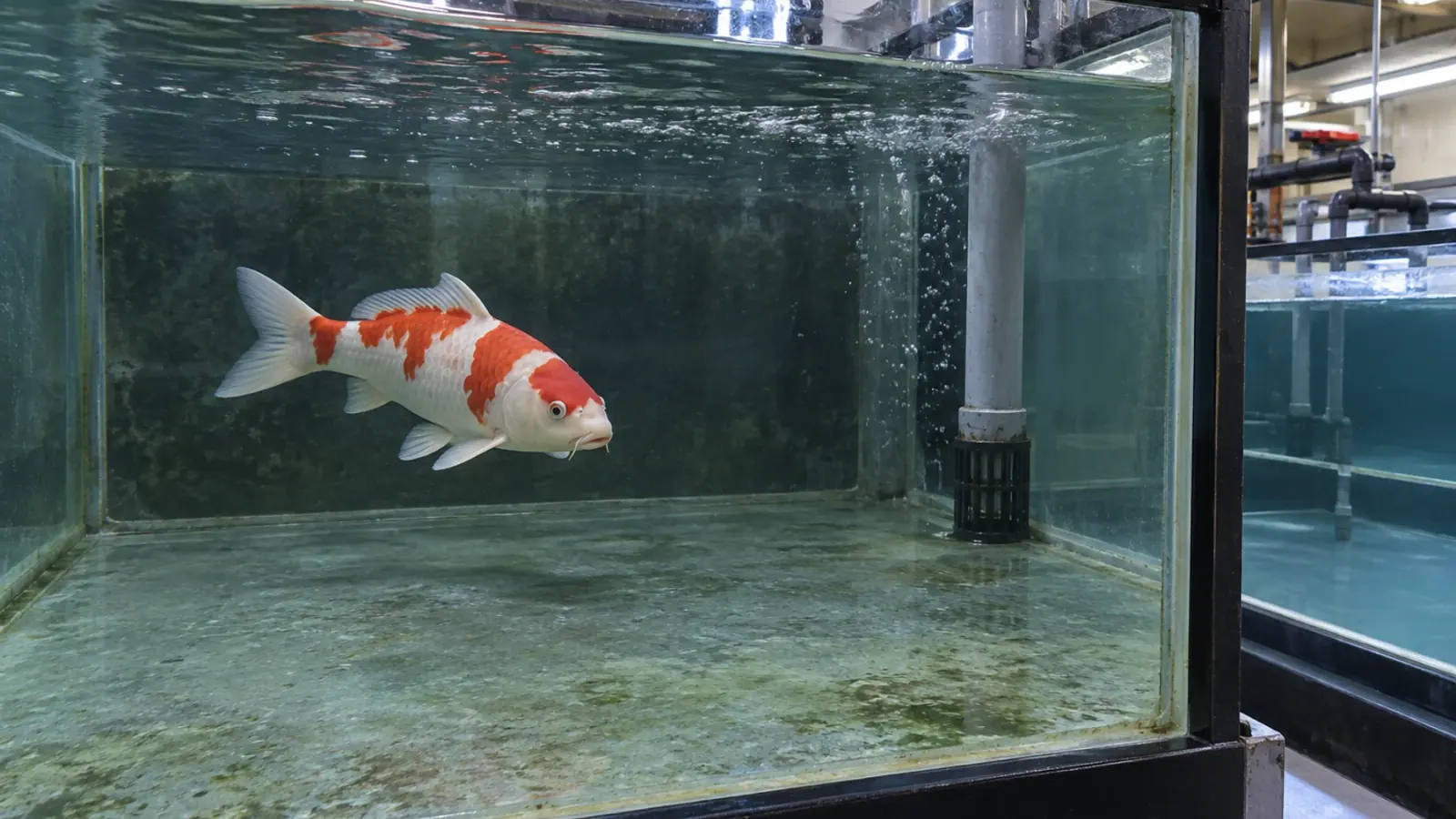
Koi coelioscopy: what can be seen, and what remains uncertain
Directly viewing a koi’s internal organs can support a diagnostic investigation, but the procedure remains invasive. A 2019 study of sixteen fish separates readily visible structures from difficult targets and documents complications that require postoperative follow-up.
- Content type
- Practical guide
- Sector
- Public aquariums
- Animal group
- Cyprinids
Start with the organ and the clinical question
Coelomic swelling or abnormal buoyancy in a koi carp, Cyprinus carpio, may prompt investigation of internal organs. Coelioscopy provides a direct view through an endoscope introduced into the body cavity through an incision. It requires anaesthesia, appropriate veterinary facilities and a plan for postoperative care.
For a professional collection, the indication depends on the suspected organ and whether viewing it would alter the next decision. Seeing an organ is not the same as determining the cause of its disease, and being able to introduce an instrument does not establish an indication for doing so.
A 2019 study by Brittany Stevens and colleagues helps define those boundaries. It examined anatomical access and procedural tolerance in predominantly healthy koi. It did not compare diagnostic strategies in a series of fish with confirmed coelomic disease.
Sixteen koi and two entry sites
The researchers studied sixteen koi approximately eighteen months old, with a mean total length of 28.2 centimetres. Eight were allocated to an approach cranial to the pelvic girdle and eight to an approach caudal to it. Entry and visualisation of selected structures were scored.
After surgery, the fish were monitored. Eight were euthanised at two weeks and eight at eight weeks for necropsy and histological assessment. This allowed the team to look for tissue changes that normal-looking swimming or feeding might not reveal. It also defines the period over which postoperative outcomes were assessed.
All fish recovered from anaesthesia and resumed feeding by the next day. That immediate recovery is useful information, but it does not summarise the longer course. Subsequent examinations identified tissue injury, adhesions and delayed wound healing.
Some organs were much easier to see
The liver, intestine and gonads were readily visible. The anterior kidney and heart could also be identified. Visualising the posterior kidney and swim bladder required more searching or manipulation. A description of coelioscopy as an equally effective examination of every internal organ would therefore be misleading.
The spleen and gallbladder were not visualised. Their position among other tissues made access difficult, and attempts to find them raised concerns about damaging fragile surrounding structures. The investigators stopped pursuing these targets. An organ that has not been seen cannot be reported as normal on the basis of that examination.
This anatomical distinction helps frame an individual case. A poorly accessible target may call for a different investigation. An accessible structure may support a focused examination when the expected information is useful. Fish size and operator experience also remain relevant to what can be achieved.
A practical preference is not a proven superior approach
Entry and visualisation scores did not differ significantly between the two approaches. With eight fish per group and a specific scoring system, absence of a significant difference does not demonstrate equivalence under all clinical conditions.
The authors found the caudal approach more convenient for viewing some dorsal and caudal structures. They recommended considering it for those targets, while acknowledging that the measured visualisation scores had not established superiority. That distinction should remain visible when the paper is used in clinical planning.
An approach should therefore be discussed in relation to the target organ and the individual fish. The study does not justify a universal entry-site rule for koi, and still less for the many other fish species kept in public aquariums.
Normal feeding did not exclude internal complications
Transient bleeding and rupture of gonadal capsules occurred during the procedures. Necropsy later revealed adhesions between viscera and the body wall. Some incision sites remained inflamed and incompletely healed at eight weeks.
Histopathology documented tissue reactions that could not be inferred from the return of appetite alone. The authors discuss water temperature and suture material as possible contributors to slow repair. These factors were not experimentally compared, so their individual contributions cannot be quantified from this trial.
Analgesia, anaesthetic monitoring and postoperative assessment belong within the veterinary plan. The drugs and procedures used for this experiment are not a prescription that can be transferred unchanged to a collection. The patient, its holding conditions and the proposed intervention determine the plan needed.
Distinguish feasibility from diagnostic performance
The findings do not establish sensitivity for detecting a tumour, infection or another disease. Fish with inflammatory coelomic disease may have more extensive adhesions than the animals studied, potentially making visualisation harder. The authors explicitly identify this limit to extrapolation.
Repeated procedures also deserve consideration. An intervention can alter tissues and affect subsequent access. Similarly, the general possibility of collecting an endoscopic biopsy does not mean this experiment validated sampling of every organ. Visual examination, obtaining a specimen and reaching a final diagnosis are separate steps.
The study documents which organs were accessible and which complications occurred under its conditions. It supports an informed discussion of the technique, rather than a guarantee that an apparently uncomplicated recovery indicates complete healing.
Plan the investigation and the follow-up together
In public aquariums, the clinical record should connect presenting signs, the intended target and postoperative monitoring criteria. Appetite, swimming, wound appearance and clinical progression provide complementary information. None is a complete measure of internal healing on its own.
Vetofish helps teams discuss indications and organise suitable diagnostic examinations. Coelioscopy is valuable when it answers a useful question for the patient; its expected reach, limitations and risks should be explicit before the examination and throughout follow-up.


