

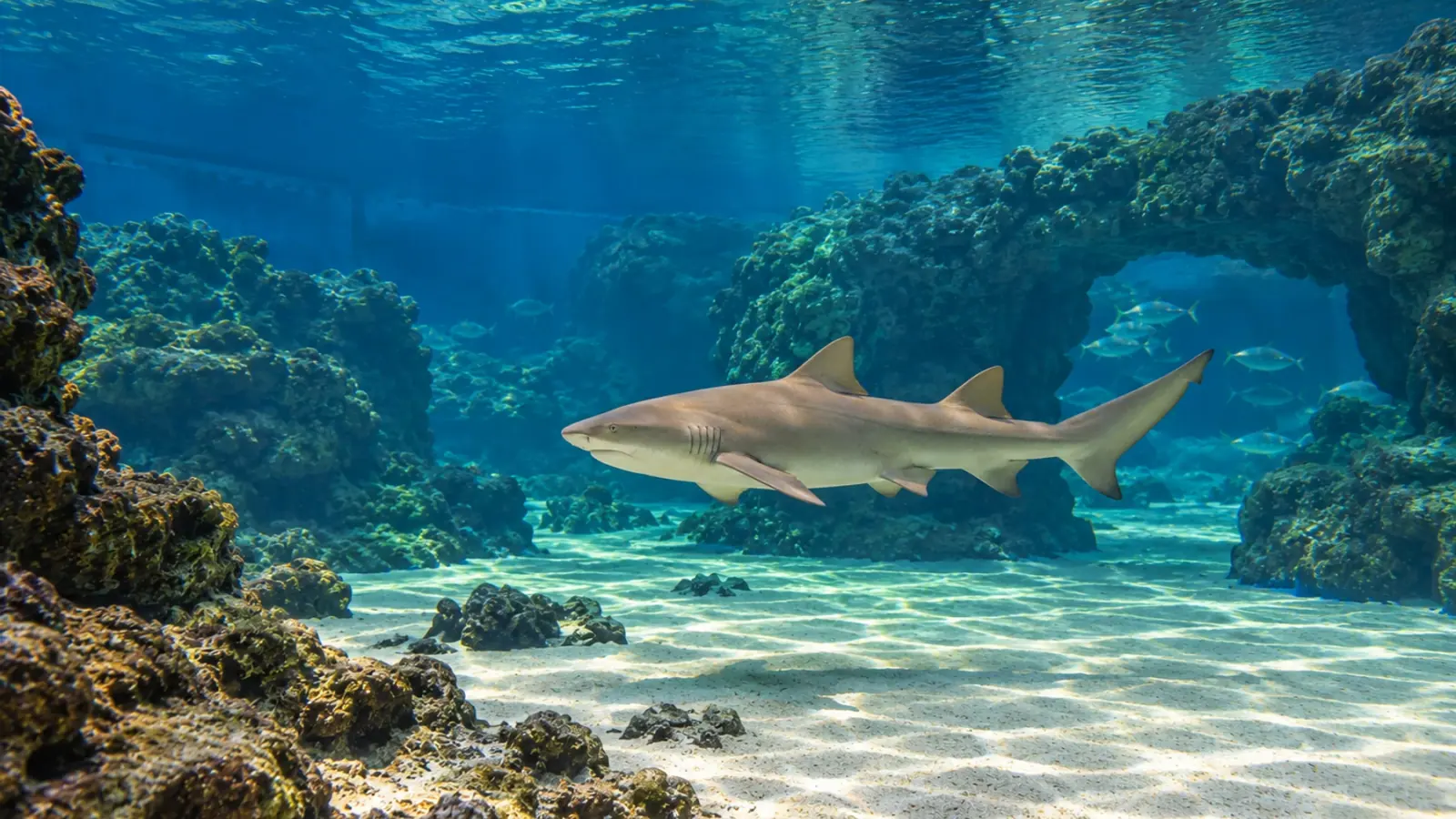
Lemon shark mouth lesions: investigate beyond the teeth
A lesion around a shark’s mouth may have little to do with its teeth. A 1997 series involving four lemon sharks illustrates why skin examination, parasite identification and shared water systems matter before treatment is chosen.
- Content type
- Disease profile
- Sector
- Public aquariums
- Animal group
- Elasmobranchs
- Theme
- DiagnosticsDiseases
- Keywords
- HistopathologyQuarantineStress
A dark or eroded area around a shark’s mouth should prompt assessment of the whole animal. Its location does not establish dental disease, and a photograph alone cannot identify its cause. In a public aquarium, the investigation needs to connect lesion descriptions, behaviour, diagnostic samples and the history of the water system.
A historical lemon shark series illustrates this approach. Poynton and colleagues reported mouth-area and skin lesions associated with a monogenean in four Negaprion brevirostris in a Florida aquarium in 1997. The report is useful as a clinical example, not as evidence of a new outbreak or a current treatment protocol.
A separate exhibit can still share exposure
Three adult lemon sharks occupied a large display with other fish. An immature female lived in a separate exhibit that shared the same water. Physical separation of the animals therefore did not provide complete separation of their aquatic environment, an important distinction when considering possible routes of exposure.
Reported signs included rubbing against rocks and walls, a dark haemorrhagic band around the mouth and irregular grey patches on the head. Some lesions produced abundant mucus. Parasites were collected from head lesions on two sharks and mouth lesions on the immature shark. Infrequent samples from the fourth animal were negative despite persistent lesions.
Morphological examination identified the worms as Neodermophthirius harkemai. The parasite was associated with the lesions, but the findings did not establish a single cause for every affected area. The three adults survived. The immature shark died, but necropsy and histopathology did not establish why. Its death should not be attributed to the parasite as a demonstrated outcome of the report.
Describe the affected tissue precisely
For a new case, establish a repeatable description before assigning a diagnosis. Record location, extent, colour, surface changes, bleeding, mucus and progression. Skin around the mouth, lips, the oral cavity and teeth are different anatomical sites. Grouping them under a general label such as “mouth disease” can obscure the question that sampling needs to answer.
Comparable photographs help document progression when angle, distance and lighting are reasonably consistent. Images should stay attached to the animal’s identity and date. Feeding observations provide additional context: altered prey capture, rejection of food or a change in feeding behaviour warrants assessment, but none identifies a specific disease by itself.
Record changes elsewhere on the body and any rubbing behaviour. In the historical series, the mouth-area lesions formed part of a broader skin problem. Restricting attention to the teeth would have overlooked the association that made a skin-parasite investigation relevant. This does not mean that every oral lesion in another shark has a parasitic origin.
Choose samples to resolve a diagnostic question
Sampling decisions should reflect the individual, the lesions and the risks of handling. A parasitological examination can look for an external parasite, while a biopsy or other laboratory tests address different possibilities. The veterinary team should agree what each result could establish before exposing the shark to restraint or an invasive procedure.
Chang and Okihiro’s 2024 guide to elasmobranch necropsy includes structured examination of the lips, gingiva, teeth and oral cavity. It provides useful anatomical context for sharks and rays. However, a post-mortem examination sequence is not a sampling protocol for a living animal, and the distinction should remain clear when planning clinical investigations.
A negative result belongs to a particular site, method and sampling occasion. In the lemon shark report, negative findings from occasional samples did not explain one animal’s continuing lesions. The authors also suspected secondary bacterial involvement, but bacteriology and histopathology had not been performed routinely. That proposed contribution remained unconfirmed and should not be presented as a diagnosed mixed infection.
Recurrence calls for a system review
The authors reported parasite removal and skin recovery after adults were transferred to an isolated facility and treated. Lesions recurred two months after return to the original display. A reservoir in the exhibit system was considered plausible, although persistence of parasites despite treatment could not be excluded completely.
The recurrence did not identify a particular carrier or prove how long parasites survived without their preferred hosts. It nevertheless provides a reason to map shared water, transfers, equipment movements and recent introductions. Animals remaining in the exhibit need assessment; proximity alone is not evidence that they are infected.
Quarantine should therefore be described through barriers and release criteria, rather than the name of a room or tank. Apparently normal skin at arrival and a previous treatment do not establish that an introduction is risk-free. Follow-up after transfer back into a collection is part of assessing whether the original problem has been controlled.
Historical interventions are not ready-made prescriptions
The report describes several interventions used in sequence, with variable results. It was a small descriptive case series without randomised treatment comparisons. Improvement after isolation and treatment does not separate every contribution of those changes, and the reported chemical practices should not be copied directly into another collection.
Species, water chemistry, filtration, other inhabitants and the applicable conditions of medicine use require a site-specific veterinary assessment. This is especially relevant in a mixed exhibit containing organisms with different treatment tolerances. Parasite identification and the wider differential diagnosis should precede selection of an intervention.
Use the mouth lesion as the beginning of the investigation
The lasting lesson is to examine the skin and accommodation system as well as the mouth, and to retain the limitations of each finding. A clinically useful record distinguishes observations, confirmed diagnoses and hypotheses that still need testing.
Through its diagnostic and laboratory service, Vetofish can help public aquariums prepare sampling plans and interpret clinical, parasitological and tissue findings together. The aim is an investigation appropriate to the shark and its collection, without attempting to diagnose a mouth lesion remotely from an image alone.


