

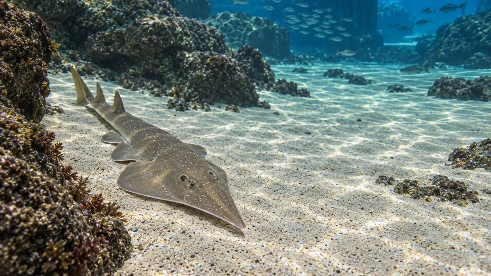
Photobacterium in guitarfish: investigating an aquarium outbreak
Three common guitarfish died rapidly in an Italian aquarium. Examination identified Photobacterium damselae subsp. damselae. The report supports a coordinated investigation, while leaving the infection source and effective treatment unresolved.


- Content type
- Scientific news
- Sector
- Public aquariums
- Animal group
- Elasmobranchs
- Theme
- DiseasesDiagnostics
- Keywords
- HistopathologyPCRStress
Respiratory distress in a guitarfish requires prompt assessment of the animal and its aquarium environment. It does not identify a single cause. Bignami and colleagues describe an episode in an Italian aquarium in which husbandry records and complementary examinations supported a systemic bacterial infection. Their report also illustrates the information that remains missing when death follows quickly and the investigation is largely post-mortem.
Three adults in a mixed-species aquarium
The episode occurred in December 2023 in three common guitarfish, Rhinobatos rhinobatos. Two females and one male, each more than twenty years old, had lived at the aquarium for over fifteen years. Their 56,000-litre tank held both cartilaginous and bony fishes. This established collection provides a different kind of evidence from a controlled infection experiment.
The guitarfish developed respiratory distress and ataxia and died within approximately one day. Such a short course leaves little opportunity for repeated clinical observations or sampling of living animals. Aquarium staff can improve the information available by maintaining a shared chronology: which individual changed, when the change began, feeding history, observed behaviour and any interventions carried out.
The authors report no recognised change in the monitored water parameters around the episode. That finding does not establish that environmental stress was absent. It means the available records did not reveal a shift that directly explained the deaths. Measurements from this particular tank should not be converted into universal husbandry limits for common guitarfish in other collections.
Complementary evidence supported systemic infection
Post-mortem examination identified haemorrhagic enteritis and lesions affecting several organs. Histopathology showed bacterial aggregates associated with affected tissues. This evidence differs from detecting an organism in tank water: it places bacteria alongside tissue damage in the animals examined. Interpreting the lesions and the laboratory findings together is essential to the investigation.
Bacteriological examination produced abundant growth with a dominant colony morphotype from organ samples. Identification combined laboratory approaches, including MALDI-TOF mass spectrometry and PCR, to characterise Photobacterium damselae subsp. damselae. The distinction from subsp. piscicida matters. Similar names do not justify treating findings for the two subspecies as diagnostically or clinically interchangeable.
The agreement between tissue findings and bacterial identification supports the septicemia diagnosis reported by the authors. It does not establish the initial entry route. Cultures still need to be interpreted with sample origin, collection quality and tissue examination. Detecting a bacterium without that context would not be enough to explain every mortality event in a mixed-species collection.
Negative findings answer particular questions
The reported parasitological examinations did not identify parasites. Testing pooled brain material for betanodavirus by RT-PCR was negative. These findings address the hypotheses examined with those methods. They do not demonstrate the absence of every viral or parasitic agent from the affected fish, their environment or the other species sharing the aquarium system.
The tissue selected, sampling time and test method affect what can be detected. An investigation record should retain those details beside the result. A statement such as “negative virology” would conceal the agent actually targeted and the material tested. That loss of detail can encourage a later reader to infer a much broader exclusion than the analysis supports.
For future incidents, the veterinarian and diagnostic laboratory can agree sample allocation, storage and transport before material is collected. Culture, histology and molecular testing have different requirements. Planning the allocation reduces the chance that preparation for one test makes another impossible, particularly when only a small amount of suitable tissue or few affected animals are available.
Long-term captivity did not reveal the infection source
A long history at an aquarium does not guarantee freedom from infectious risk. It does provide a timeline to investigate. Recent animal movements, equipment sharing, feeding and interventions can be compared with the onset of signs. A plausible pathway remains a hypothesis unless evidence connects it to the event. Familiarity with the animals should not replace that reconstruction.
The authors discuss stress as a possible contributor without establishing its cause in these cases. The report therefore cannot justify assigning responsibility to a particular handling event or item of equipment. Keep observations, proposed mechanisms and missing information distinct. This makes it easier for the team to investigate alternatives and review the explanation when further results become available.
Surveillance of remaining animals can use signs defined with the clinical team, including respiration, swimming, posture and feeding changes. An observed change should trigger proportionate assessment rather than an assumption that every tank inhabitant has the same infection. Individual records and observations of the group complement each other; neither replaces a clinical examination when one is needed.
A case report is not a treatment comparison
Three animals from one facility cannot establish how frequently the infection occurs in common guitarfish or compare treatment strategies. The report does not demonstrate the effectiveness of an antibiotic, vaccine or water adjustment for this species. Findings from other fish should not be converted into an elasmobranch prescription without a separate assessment of their applicability.
The case is still useful for reviewing operational readiness: veterinary contact, sample transport, access to historical records and responsibility for bringing the findings together. Measures following an investigation should address identified risks. System-wide disinfection or treatment based solely on the name of a bacterium may miss the relevant pathway and create additional management problems. The decision needs the full clinical and environmental context.
Conclusion and Vetofish support
This report documents systemic infection in three common guitarfish while leaving the source and effective treatment unresolved. Our animal health expertise can help public aquarium teams prepare diagnostic investigations, bring tank records together with laboratory findings and define surveillance suited to the animals in their care.


