

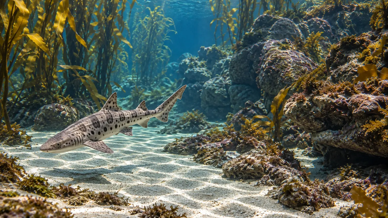
Shark and ray necropsy: why the head needs attention
A California case series shows why shark and ray post-mortems should include the head, inner ears and olfactory structures. The approach complements a whole-body examination and depends on suitable samples rather than a visible lesion alone.
- Content type
- Protocol / technique
- Sector
- Public aquariums
- Animal group
- Elasmobranchs
- Theme
- DiagnosticsDiseases
- Keywords
- HistopathologyPCRBiosecurity
Include the head in the diagnostic plan
A shark or ray post-mortem should connect the animal’s history with lesions and laboratory findings. The head deserves specific attention because the sensory structures and brain can contain changes that an external examination will miss. The chondrocranium is the cartilaginous braincase. Its anatomy requires an approach suited to elasmobranchs rather than one copied from a bony mammalian skull.
Chang and Okihiro described such an approach in 2024, drawing on work with stranded wild animals and aquarium-housed elasmobranchs in California. Their protocol was developed and used in the examination of 189 animals representing 18 species between 2011 and 2023. This provides practical diagnostic experience. It does not establish disease frequencies across public aquarium populations.
Follow the relevant anatomy
The authors emphasise the endolymphatic pores and ducts, inner ears, brain and olfactory structures. Endolymphatic ducts provide a connection between the external environment and the inner ear. Examining these structures can help interpret lesion distribution and possible entry routes for particular pathogens. Recognising the normal anatomy is therefore part of choosing where to sample.
This does not make unusual swimming or loss of balance proof of infection. Behaviour observed before death remains clinical information that needs interpretation alongside other findings. A description of when the behaviour started, its progression and accompanying changes is more useful than retrospectively labelling it a brain lesion. Several possible explanations may remain until the investigation is complete.
Agree on samples before dissection
The published approach combines gross examination, cytology, microbiology, histopathology and molecular methods where appropriate. These tools answer different questions. Identifying an organism, describing an inflammatory response and showing an association between the two should not be treated as equivalent results. Diagnostic confidence comes from their relationship to the case as a whole.
Preparation should involve the veterinarian and receiving laboratory before tissues are collected. Sampling sites, containers and storage requirements depend on the intended tests. Material placed in a fixative cannot simply be used for every other investigation. Submission records should preserve species, animal identity, relevant dates, storage history and any known previous treatment. Missing information can limit interpretation even when the sample arrives intact.
Protect the interpretation of microbiology
The protocol uses an aseptic approach when accessing selected cranial sites. This reduces external contamination during collection, which matters when a culture result is compared with tissue changes. A positive culture by itself does not explain how the animal became ill or establish that the organism caused its death.
The authors describe bacterial and protozoal disease in their material. The meaning of a finding depends on species, sampling site, carcass condition and agreement between tests. Negative results can also be constrained by the location or quality of the sample. Discussion between the clinician, pathologist and laboratory is more informative than treating the detected organism’s name as a complete diagnosis.
Record the post-mortem interval honestly
Autolysis is tissue breakdown after death. It can alter lesions and make organisms harder to recognise. Chang and Okihiro favour examination as soon as possible and discuss refrigeration or freezing when work must be delayed. These options involve different compromises for subsequent tests, so storage decisions should be agreed with the diagnostic team.
The aquarium can preserve essential information immediately: when the body was found, when the animal was last seen alive and how it was stored. These are three different records, not interchangeable estimates of the time of death. Photographs taken before destructive handling can retain external findings that transport or examination may later change. They also help connect clinical observations with the post-mortem record.
Keep examining the whole animal
Prioritising the head does not replace a complete necropsy. Gills, heart, digestive tract and other organs may reveal an additional or alternative explanation. The paper explicitly places cranial examination within a whole-body investigation. A striking lesion should therefore not automatically end the search for other relevant changes.
The system history matters too. Appetite, behaviour, water conditions, recent introductions and technical interventions provide context for the findings. They should be used to test explanations, rather than assign a cause in advance. When animals share a water circuit, the health assessment should consider that connection without assuming that every individual is infected or that whole-system medication is warranted.
Transfer the method without transferring a prevalence estimate
The Californian material includes different circumstances, notably strandings. The animals were not randomly sampled from healthy wild or captive populations. Results cannot therefore be converted into a universal risk estimate for sharks and rays in European aquariums. The most transferable contribution is the anatomical approach and the planned combination of diagnostic methods.
The authors also explain that expanded mortality data are intended for later publication. Readers should retain the evidence actually presented: anatomical landmarks, sampling methods and examples supporting diagnostic interpretation. These findings do not justify a general ranking of causes of death for every elasmobranch collection.
Make the examination useful for the collection
A useful necropsy begins before the carcass is opened. Planning samples, preserving identity and recording storage conditions help the team interpret lesions alongside laboratory results and husbandry history. Even a carefully conducted investigation may leave uncertainty, which should remain explicit in the report and subsequent management decisions.
Our diagnostic and laboratory service can help public aquariums plan sampling, discuss findings with laboratories and consider proportionate next steps for the collection. A diagnosis should inform those steps rather than be assumed in advance.


