

Can tank water reveal every zebrafish pathogen?
Water and detritus can reveal some agents without sacrificing fish. An open study in recirculating systems also shows which pathogens environmental testing alone may miss.


- Content type
- Scientific news
- Sector
- Research facilities
- Animal group
- Zebrafish
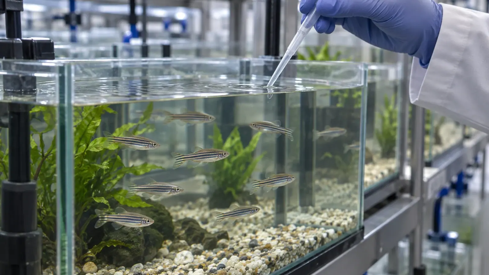
Collecting water from a zebrafish tank is less invasive than sacrificing an animal for tissue analysis. The option is attractive to research facilities, yet a negative water PCR result does not necessarily describe the health of every fish. An open study in recirculating systems compared diseased fish with water and detritus collected from their tanks. Its practical lesson is to select samples according to the pathogens of interest and the decision the test must support.
A comparison in recirculating systems
Miller and colleagues examined Danio rerio with clinical signs in a facility operating several recirculating water circuits. They compared real-time PCR results from fish with results from tank water mixed with detritus and concentrated on a membrane. Their question was whether an environmental sample could usefully complement testing of affected animals. The protocol did not establish a universal sensitivity for water PCR across all tanks, agents or infection stages.
A positive quantitative PCR result detects the molecular target specified by the assay. On its own, it does not show that the organism is still viable or that it caused illness in a particular fish. A negative result may reflect limited shedding, too little material, the wrong collection site or treatment of water before sampling. Facilities need to define their surveillance and exclusion lists before choosing a convenient sample type.
Some agents appear mainly in fish
In this study, Pseudoloma neurophilia and Myxidium streisingeri were found exclusively or mainly in the fish examined, whereas mycobacteria were predominantly detected in environmental material. The profiles were therefore not interchangeable. Water testing that helps detect one agent may miss another present in the tissues of diseased animals. The authors concluded that both sample types were needed to cover a wider range of agents in their setting.
Biology and sampling method both matter. Pathogens occupy different tissues, leave their hosts at different rates and persist differently in water or detritus. PCR on a fish selected because of its clinical signs answers a different question from PCR on a mixture of water and particles. Labelling both results simply as a negative health status would conceal the population and compartment actually tested.
Make environmental sampling traceable
Recirculating water may connect several tanks, but the population represented by a sample depends on the collection point, flow, filtration and disinfection. In the study, water and detritus came from tanks holding fish with clinical signs. That targeted design does not automatically validate sampling an entire circuit or an apparently healthy colony. A local protocol should record the volume, location, collection time, membrane, storage conditions and contamination controls.
Detritus provides another view of the tank, but its accumulation period changes with cleaning. Teams comparing results over time should record maintenance, flow changes and equipment modifications. In quarantine, a result should be reported for the fish and tanks actually represented. Water from a shared circuit cannot by itself clear every neighbouring genetic line. The report should also identify any samples that could not be collected, instead of treating missing material as a negative test.
That distinction matters when a research group receives animals from another institution. A report based on a tank containing sick fish, a routine circuit sample and a sample from a newly arrived quarantine group have different purposes. Each should identify the collection date, affected or apparently healthy population, pathogens on the assay panel and the point where water was drawn. If animals have moved between circuits, that history belongs beside the test result. The receiving team can then judge whether additional observation or targeted testing is needed, rather than inheriting an unexplained label of “PCR negative.”
When do fish still need examination?
Water and detritus can guide routine surveillance and reduce some animal sampling. Fish showing signs remain a separate diagnostic source. Depending on the question, clinical examination, tissue testing and histopathology may be required to connect a molecular signal with lesions and disease. Before testing, a facility should define which signs trigger fish examination, which targets are sought and how disagreement between sample types will be investigated.
Sampling biosecurity affects the credibility of results. Dedicated equipment, a planned order of movement between rooms, clear labels and laboratory controls help prevent material from one circuit reaching another sample. Read results alongside introductions, mortality, clinical observations and interventions. A single detection without those records can lead either to an unnecessarily broad response or to misplaced confidence.
Reduce sacrifices while preserving diagnosis
The study concerned one facility, clinically affected fish and a defined group of pathogens. It does not demonstrate that environmental samples replace sentinels everywhere or that another circuit will yield the same pattern. Reducing animal use must be assessed together with surveillance quality: a noninvasive test is useful when it answers the real question and the circumstances requiring examination of fish remain explicit.
For research facilities, our diagnostic and analysis service can help select samples by pathogen and explain the limits of a negative result when fish move between facilities.


