

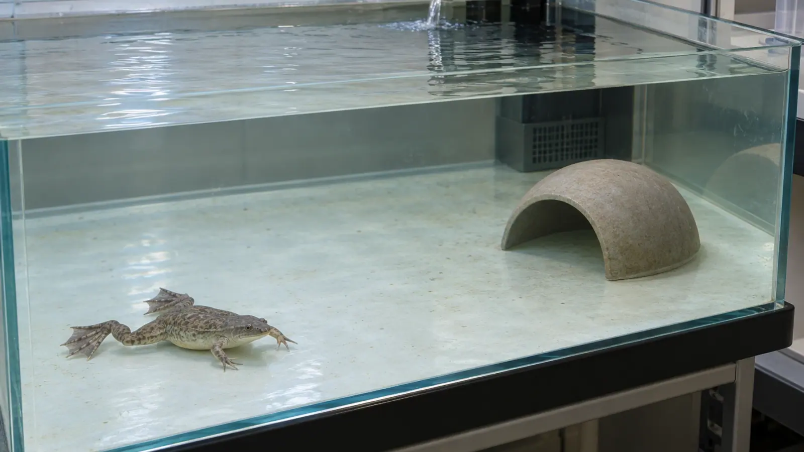
Xenopus skin disease: investigating cutaneous capillariasis
Unusual skin shedding in Xenopus laevis can accompany cutaneous capillariasis. Shed skin and histology can contribute to the diagnostic investigation; redness or a bacterial culture alone does not identify the underlying cause.
- Content type
- Disease profile
- Sector
- Research facilities
- Animal group
- Xenopus
- Theme
- DiseasesDiagnostics
Skin fragments on the bottom of a Xenopus tank are not, on their own, a parasitic diagnosis. Shedding is part of the animal’s biology. Unusual shedding accompanied by rough skin, weight loss or reduced activity, however, warrants investigation. Cutaneous capillariasis is one of the conditions to consider in Xenopus laevis.
The parasite is generally called Pseudocapillaroides xenopi. Older reports also use Capillaria xenopodis and related name combinations. Retaining these synonyms when searching historical records helps locate relevant cases. This skin nematode should not be confused with Pseudocapillaria tomentosa, the intestinal parasite of zebrafish.
The parasite occupies the epidermis
Armed Forces Institute of Pathology case records describe nematodes and eggs within tunnels in the epidermis. Hyperplasia means an increase in the number of cells in the tissue. It can occur alongside structural changes and inflammation, compromising the skin barrier.
Amphibian skin contributes to exchanges with the surrounding water. Extensive lesions are therefore more than a cosmetic change. The institutional descriptions connect epidermal damage with possible disruption of skin function and secondary infection. The clinical importance needs assessing in the animal concerned, rather than from a photograph alone.
The reported life cycle is direct, without a necessary intermediate host, with developmental stages present in the epidermis. This makes the group and shared equipment relevant to an investigation. It does not date the introduction of infection or identify the operation responsible for transmission in an individual case.
Two reports illustrate different presentations
An April 2009 case presentation concerned females used for cell-cycle research. Five dead frogs were submitted for examination. All five had the cutaneous infestation, while cultures from skin and cardiac blood were negative. This number describes the animals examined; it is not a prevalence estimate for the whole colony.
A December 1999 case described parasitic dermatitis with Aeromonas cultured from external lesions. Together, the reports illustrate situations in which secondary bacterial involvement was recorded or was not recovered by culture. A bacterial culture does not take the place of examination for the parasite.
These are historical case records. They inform a continuing differential diagnosis, without demonstrating a new outbreak in present-day research facilities or providing a current estimate of infection frequency. A colony health plan needs to retain these limits when using the case descriptions.
Give shed skin a defined place in the investigation
The diagnostic descriptions include direct examination of shed skin fragments and histopathology. Collected skin can therefore be useful investigative material when its origin and condition are known. An old fragment recovered from a shared circuit does not have the same attribution as material collected from an identified holding tank.
The receiving laboratory can specify the appropriate container and preservation method before collection. Material intended for direct observation and tissue fixed for histology are not interchangeable. The submission should identify the tank, collection date, animals affected, observed signs and any treatment already given.
Failure to find a parasite in one fragment cannot establish that a group is free of infection. Lesion distribution, the amount of material examined and preservation quality influence the examination. If signs persist, the veterinarian and laboratory can select proportionate additional investigations while considering the risks of handling the animals.
Investigate contacts as well as the individual
Observations need to be connected with group history: origin, recent introductions, transfers, linked tanks and shared equipment. Quarantine arrangements and movement of equipment are relevant biosecurity questions. Their purpose is to identify possible contacts, without assigning blame to a supplier on the timing of a delivery alone.
The team should also follow appetite, activity, skin changes and water conditions. An environmental problem can coexist with parasitism. Correcting an abnormal water parameter does not identify an infectious agent, just as a positive parasitological finding does not automatically explain every sign in the group.
Animals deteriorating clinically need timely assessment. Care and reassessment criteria should be agreed with the responsible veterinarian. Collecting a planned series of samples should not postpone intervention in an animal that needs attention. The investigation and animal-care decisions need to remain connected throughout.
A case report is not a universal treatment schedule
Historical records mention anthelmintic treatment. They do not provide a generally applicable protocol for another colony, housing system or compromised animal. Drug selection, route and administration arrangements require veterinary assessment of the animals and their holding system.
Diagnostic confirmation also needs separating from follow-up after intervention. Agreeing what will be monitored in advance allows clinical progress and laboratory findings to be reviewed together. One negative observation after treatment should not be treated as certain eradication from every linked tank.
The record should preserve which animals and sites were assessed, when they were examined and how the results were obtained. This makes a later comparison interpretable even when staff, tanks or laboratory methods change. A brief statement that a group tested negative loses much of this information.
Link skin findings with group surveillance
Cutaneous capillariasis illustrates why skin and shed material deserve examination when a Xenopus colony develops unexplained problems. Redness, a positive bacterial culture or one negative skin fragment each provides partial information. Their relationship with the lesions and group history guides the next decision.
Vetofish’s diagnostic and laboratory service can help identify suitable submission material and interpret findings. For research facilities, support can connect the individual case, contacts between tanks and a surveillance plan appropriate to Xenopus.


