

Red-leg in Xenopus: investigate beyond skin redness
Ventral redness in Xenopus laevis does not establish Aeromonas infection. Describing signs, reviewing husbandry and interpreting lesions alongside microbiology helps avoid a diagnosis based on appearance alone.
- Content type
- Disease profile
- Sector
- Research
- Animal group
- Xenopus
- Theme
- DiseasesDiagnostics
Red skin is an observation, rather than an identified cause
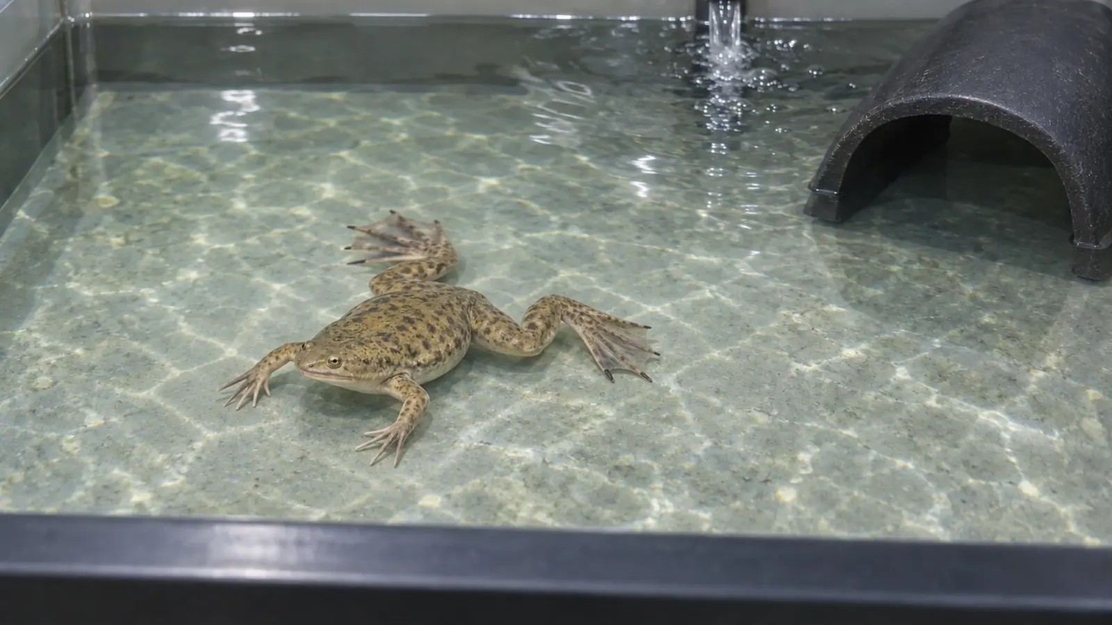
Ventral or limb redness, reduced appetite and altered behaviour in an African clawed frog warrant assessment. The term “red-leg” has historically been associated with bacterial dermatosepticaemia. It does not identify a causative organism from the appearance of the skin, and should not be treated as a diagnosis established by a photograph.
In their review of amphibian diseases, Densmore and Green emphasise that this syndrome has frequently been overdiagnosed. Different conditions can look similar, while bacteria recovered after death may not explain the original disease. For a research facility housing Xenopus laevis, that distinction affects how an alert is described, how examinations are prepared and how findings are interpreted.
Keep observations separate from hypotheses
Erythema means redness of the skin. Record where it occurs, its extent and how it changes, without turning that description into proof of systemic bacterial disease. Lesions, swelling or loss of appetite add clinical information, but none of these signs identifies a particular bacterium by itself.
The review makes clear that red-leg syndrome is not synonymous with Aeromonas hydrophila infection. Other bacteria have been associated with comparable presentations, and viral or fungal infections may enter the differential diagnosis. This is a reason for veterinary investigation, rather than selecting treatment from the visible colour of an animal.
A useful record distinguishes what was observed, what is suspected and what has been demonstrated. “Ventral redness observed” belongs to the first category; “suspected bacterial dermatosepticaemia” belongs to the second. A final diagnosis requires findings to be interpreted together. Keeping these categories explicit prevents an early hypothesis from becoming an accepted fact simply through repetition during handovers.
This distinction also matters when information reaches researchers who have not examined the animals. A concise update should carry the degree of certainty with the finding. A provisional description should remain provisional in study records and subsequent reports.
Place each animal within its husbandry history
Densmore and Green stress the importance of husbandry and environmental management in disease among captive amphibians. These factors may contribute directly or indirectly to a clinical problem. Their relevance does not establish that a specific husbandry failure occurred, or remove the need to investigate other explanations.
Before discussion with the veterinarian, staff can assemble tank identifiers, the number of affected animals and a timeline. Recent transfers, handling, water changes, technical interventions and feeding observations help locate the event within daily operations. Record the time of death as accurately as possible, marking estimates clearly.
Water results need their sampling time and location. Measurements taken after corrective action do not necessarily describe earlier exposure. Similarly, one affected frog in a shared circuit does not demonstrate that every animal is infected. The extent of surveillance and biosecurity measures should follow the risk assessment, rather than an assumption about the entire population.
Records should preserve observations from unaffected tanks as well when these are available. Such information may help describe the distribution of the problem. It cannot, on its own, establish that a particular shared factor caused disease or that a separate tank is free of risk.
Understand the limits of a positive culture
The review explains that post-mortem changes can occur rapidly in aquatic vertebrates. Microorganisms may invade tissues after death, complicating interpretation. Some bacteria can also be recovered from apparently healthy amphibians. A positive culture therefore contributes evidence whose significance depends on the animal, the sample and its handling.
Histopathology can reveal tissue lesions and their distribution. Interpreting it alongside gross examination and microbiology gives a stronger diagnostic basis than any isolated result. Each method still has limits, and a negative finding does not exclude every possible disease process.
The laboratory and veterinarian should specify which examinations can address the working hypotheses. Facility staff can prepare the required history and traceability information. Sampling and preservation arrangements depend on the intended analyses; one improvised method for every specimen can reduce diagnostic value.
Protect welfare and document consequences for the study
Clinical decisions and animal welfare take priority over preserving an uninterrupted experimental series. Responsible staff need timely information so that observations, veterinary decisions and any implications for the project are recorded. Treatment selection requires veterinary assessment of the animal and the findings available.
Separate actions taken for the animals from changes made to the experiment. Altered housing, an interrupted procedure or treatment may affect scientific observations. Recording these events cannot remove every resulting limitation, but it allows them to remain visible when the study is interpreted.
A shared handover can identify the observation to follow, the person to contact and the next review point. Its purpose is to keep the care team, veterinarian and researchers working from the same information; it does not replace examinations.
Limitations and conclusion
The 2007 review concerns amphibians beyond Xenopus laevis. It does not establish current disease frequencies in French research facilities or validate a diagnostic algorithm for every colony. Its discussion of differential diagnoses and culture limitations remains useful, while assessment must address the present animals and conditions.
The practical message is to document redness and associated signs, then seek an explanation consistent with the environment, lesions and examinations. “Red-leg” can communicate an alert, but should not close the investigation.
Vetofish support
Our testing and diagnostic service can help organise examinations and interpret results with the veterinary team. Through our research facility expertise, Vetofish can support a review grounded in observations, tank history and the actual constraints of the project.


