

Cryptocaryon: examine the gills even without white spots
Few or no white spots do not rule out cryptocaryonosis. Observations published in 2026 from Primorsky Aquarium describe rapidly developing gill disease and show why gill examination matters when marine fish develop abnormal breathing.
- Content type
- Scientific news
- Sector
- Public aquariums
- Animal group
- Fish
- Theme
- DiagnosticsDiseases
Gill disease in a public aquarium setting
Stepankov’s 2026 paper brings together observations made since 2021 at Primorsky Aquarium in Vladivostok, in display and quarantine systems. It distinguishes familiar skin presentations of cryptocaryonosis from cases dominated by gill involvement. The work is descriptive: it gives no overall monitored population from which infection prevalence or comparative species risks could be calculated.
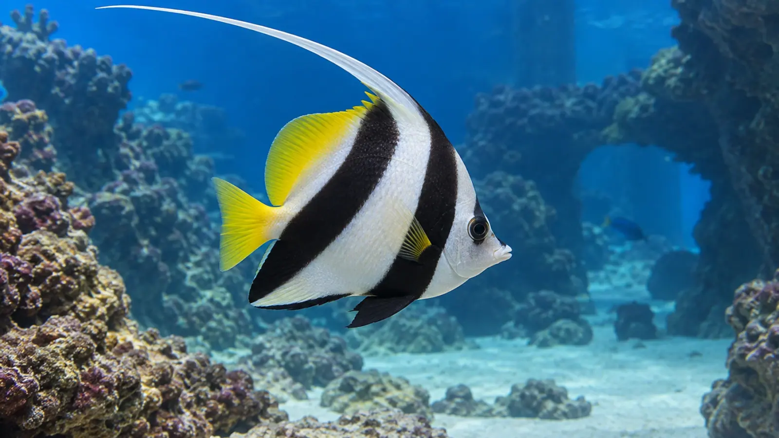
The organism discussed, Cryptocaryon irritans, is a ciliated protozoan parasite of marine fish. Skin lesions are illustrated in Chaetodon rafflesii, while gill observations include Heniochus acuminatus and Pterocaesio digramma. These examples show different presentations; they do not demonstrate an identical course in every marine species.
In the skin-dominated cases, whitish spots or nodules on the body and fins provided a visible clue. Gill-dominated cases could have few external signs while breathing deteriorated rapidly. Aquarium staff should record this mismatch between body appearance and respiratory condition.
White spots reveal only part of the problem
Gills provide the surface for gas exchange with the water. Damage there can become serious even when the flanks and fins look relatively unremarkable. Increased ventilation, respiratory difficulty or a change in position within the tank needs to be considered as part of the clinical assessment.
In the cases described, gill disease was associated with heavy parasite burdens and rapid progression, sometimes ending in death. The report does not establish a universal timetable or a prognosis for every species. It also cannot provide a numerical comparison of mortality risk between fish with and without visible spots.
The absence of white spots therefore does not exclude this parasite. It does not confirm it either. Other agents and water problems can produce abnormal breathing. Selecting a single explanation before examining the fish and system would narrow the investigation too early.
The practical implication is to record respiratory signs as carefully as skin appearance. A fish that looks clear from a distance may still need prompt assessment, while one carrying spots may have additional problems that visual inspection cannot resolve.
What a wet mount can establish
The paper uses light microscopy of native preparations from skin and gill samples. Wet mounts allow an observer to assess organisms, morphology and movement in the material collected. Their value depends on sampling location, preparation quality and the interval before examination.
A skin mucus sample and a gill sample do not answer exactly the same question. Where respiratory disease is suspected, a negative preparation from the skin alone cannot exclude organisms on the gills. Sampling must also account for the fish’s condition. Repeated handling without a clear diagnostic question can add disturbance without improving interpretation.
The study does not report molecular confirmation or a histological investigation of the lesions. Statements about tissue responses need to be read within that methodological limit. Seeing organisms at a gill surface does not by itself establish the depth of damage or every mechanism involved.
These boundaries matter when a descriptive paper is used to design a local diagnostic pathway. It supports looking beyond the skin, but it does not make every respiratory case a confirmed diagnosis of cryptocaryonosis.
Examine the water alongside the fish
Dissolved oxygen and other relevant water measurements belong alongside the health investigation. Finding a parasite is not a reason to postpone checking the system. Equally, a satisfactory measurement at one moment cannot rule out damaged gills or an earlier water event that was not recorded.
Temperature, salinity, water circulation, recent changes and the distribution of signs among fish help build a differential diagnosis. A single affected individual presents a different pattern from a problem involving several species or connected tanks. Keeping those observations together helps the veterinarian distinguish plausible explanations.
Useful referral information includes when signs began, recent transfers, previous interventions and the exact sampling sites. A microscopy result can then be linked to the condition of the fish rather than read in isolation. Results obtained before and after an intervention should not automatically be treated as equivalent observations.
A different presentation does not prove a new strain
The skin and gill forms described do not demonstrate a new species or strain of C. irritans. Location, observed size and speed of progression cannot establish genetic identity on their own. Suggestions in the paper about aspects of development require further investigation.
That limitation leaves the clinical observation useful. It directs attention towards the tissue that may need examining, without turning a descriptive report into a new classification or proof of an unfamiliar life cycle. The practical question is what evidence is needed to understand the affected fish.
Nor is the publication a comparative treatment trial. It does not provide grounds here to select a drug, concentration or treatment duration. Such a decision requires assessment of the system, species present, diagnostic findings and the establishment’s care arrangements.
Prepare a workable surveillance response
For a public aquarium, observation should include breathing and behaviour even where external lesions are inconspicuous. Quarantine records and separation of equipment support biosecurity. They do not turn a clear external examination into evidence that a fish is free of infection.
Teams can agree in advance which observations to collect and how samples will be obtained with their veterinarian. Microscopy is more informative when it answers a defined question than when it is simply added to an undirected checklist.
Vetofish helps organise laboratory testing and diagnosis for gill problems by connecting fish examination, water conditions and available methods. Respiratory condition remains the starting point: waiting for white spots may delay an examination that is already warranted.


