

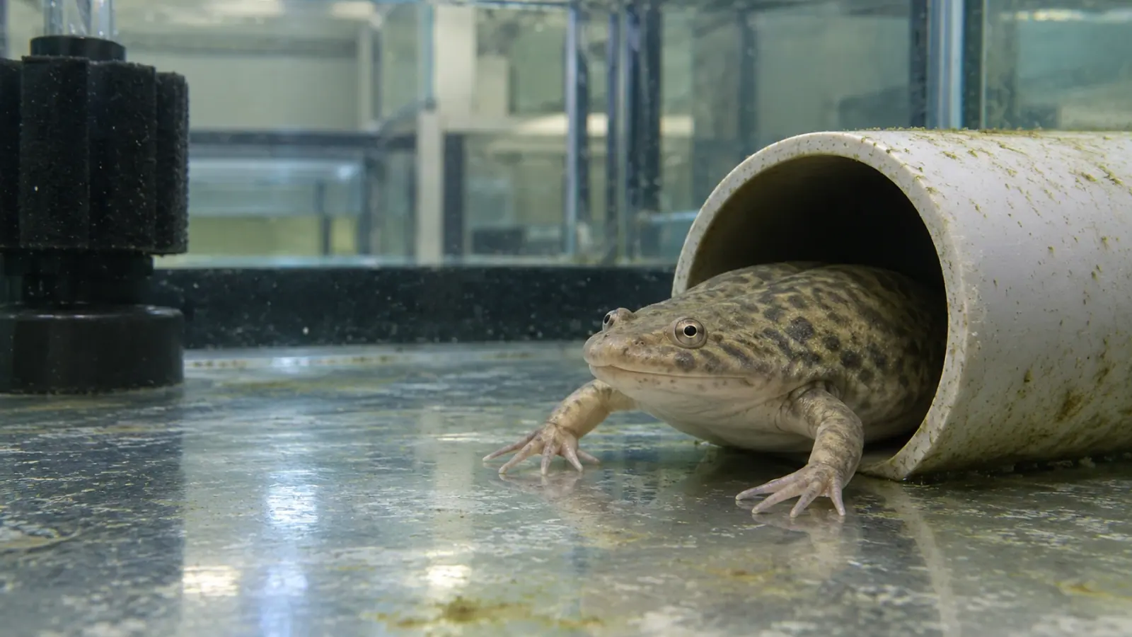
Xenopus faecal testing: protect the value of the sample
Poorly identified or delayed faecal samples can lose diagnostic value in Xenopus colonies. An institutional amphibian health manual published in 2017 helps distinguish the uses of wet mounts and concentration methods, without treating every observed organism as a cause of disease.
- Content type
- Practical guide
- Sector
- Research facilities
- Animal group
- Xenopus
Applying an amphibian colony manual to Xenopus
The 2017 manual edited by Pessier and Mendelson includes a section on testing for parasites in faeces. Its intended setting is amphibian survival assurance colonies and reintroduction programmes. It offers a useful framework for a Xenopus research facility, but it is not a validation trial carried out specifically in Xenopus laevis.
The setting limits what can be inferred for a research colony. The manual supplies neither parasite prevalence for research colonies nor a universal intervention threshold. It does not experimentally compare every test in Xenopus. Its practical contribution is to connect the organism being sought with the examination method and the condition of the sample.
Faecal examination can provide information without taking a tissue sample. It nevertheless examines particular material collected at a particular time. What it can answer depends on the question asked, as well as on what happens to be visible under the microscope.
Establish the sample’s origin before it disperses
In an aquatic tank, faeces can fragment or mix with debris on the floor. Material of uncertain origin cannot reliably identify which animal produced a finding. Collection records should retain individual identity where it is known, or explicitly state that the sample represents a tank.
Collection time, exposure to tank water, storage and transport delay also affect interpretation. Results obtained after very different delays may reflect differences in sample condition rather than a change in colony health. These details are worth recording before the material reaches the laboratory.
The manual allows pooling material from the same enclosure when the amount available is insufficient. A pooled preparation then answers a group-level question. It cannot measure the burden in a particular frog. Combining material from different tanks without distinction would lose information needed to investigate how an organism is distributed.
Collection should be planned with the animal-care team and respect normal housing and welfare. Obtaining a sample is not a reason for repeated unplanned handling or improvised changes in husbandry. The collection method needs to fit the animals as well as the laboratory question.
Fresh material supports wet-mount examination
A direct smear of very fresh faeces can reveal motile protozoa and some nematode larvae. Movement can help guide identification. It can also disappear with delay or unsuitable storage, so an organism that no longer moves was not necessarily absent or inactive when the sample was produced.
Agree the examination window and storage conditions with the laboratory before collecting. Fixing all the material at once prevents an examination that depends on living organisms. Separate portions may be needed where several methods are planned, following the receiving laboratory’s instructions.
Direct smears also have limited sensitivity for small numbers of eggs or oocysts, and debris can obscure the preparation. A negative result means the sought structures were not seen in that preparation. It does not establish that an entire tank or colony is free of infection.
This distinction helps when follow-up results appear inconsistent. Before assuming that an organism has disappeared or returned, compare the sample identities, quantities, delays and methods. A change in collection can change what is detected.
Choose concentration methods for the target
Flotation concentrates certain eggs and oocysts using the properties of the chosen medium. Sedimentation offers a complementary approach for heavier material, including some trematode eggs. The manual also describes the Baermann method for recovering living larvae.
These methods are not interchangeable. Selection depends on the suspected organism and life stage, the quantity available and laboratory expertise. Asking for a “complete faecal test” without explaining what needs investigating leaves an important part of the decision unresolved.
Faeces do not reveal only organisms whose adults live in the intestinal lumen. The manual notes that larvae of parasites occupying another organ at the adult stage can be detected through this route. Finding an organism in faeces therefore does not prove that all its stages are intestinal or that it invades the gut wall.
No single preparation establishes the complete biological relationship between an organism and its host. Its location in the submitted material is one observation to interpret alongside other evidence.
Presence does not automatically establish disease
Ciliates, opalinids and several flagellates can occur in amphibians without necessarily causing illness. Opalinids should not be classified as true ciliates merely from their surface appearance. Reliable identification requires more than noticing movement in a quick examination.
Commensalism is a different relationship from harmful parasitism. Interpretation should consider body condition, clinical progress, any digestive signs and husbandry circumstances. An unusually abundant organism may warrant investigation, but abundance alone does not provide a validated treatment threshold for Xenopus.
Poor condition or mortality may require additional investigations. Histopathology can document tissue lesions when that is the question, whereas faecal examination primarily describes material being shed. The methods address different aspects of a problem; their combination should be chosen deliberately rather than assumed to answer everything.
This also prevents a positive faecal result from becoming an unsupported explanation for unrelated signs. An organism can be present while another process accounts for the animal’s decline. Establishing a cause requires the relevant clinical and pathological evidence.
Make the result usable for the colony
In a research facility, a shared collection procedure makes observations easier to compare over time. Tank identity, collection conditions, requested method and previous interventions should remain linked. These records are useful both for quarantine arrivals and for an investigation within an established colony.
Biosecurity includes collection equipment and its movement between tanks. It complements diagnostic testing without making a negative preparation a certificate of freedom from every agent.
Vetofish supports laboratory testing and diagnosis for Xenopus colonies. Defining the question with the laboratory before collection preserves what the sample can still reveal, then allows the next decision to be based on results whose limits are understood.


