

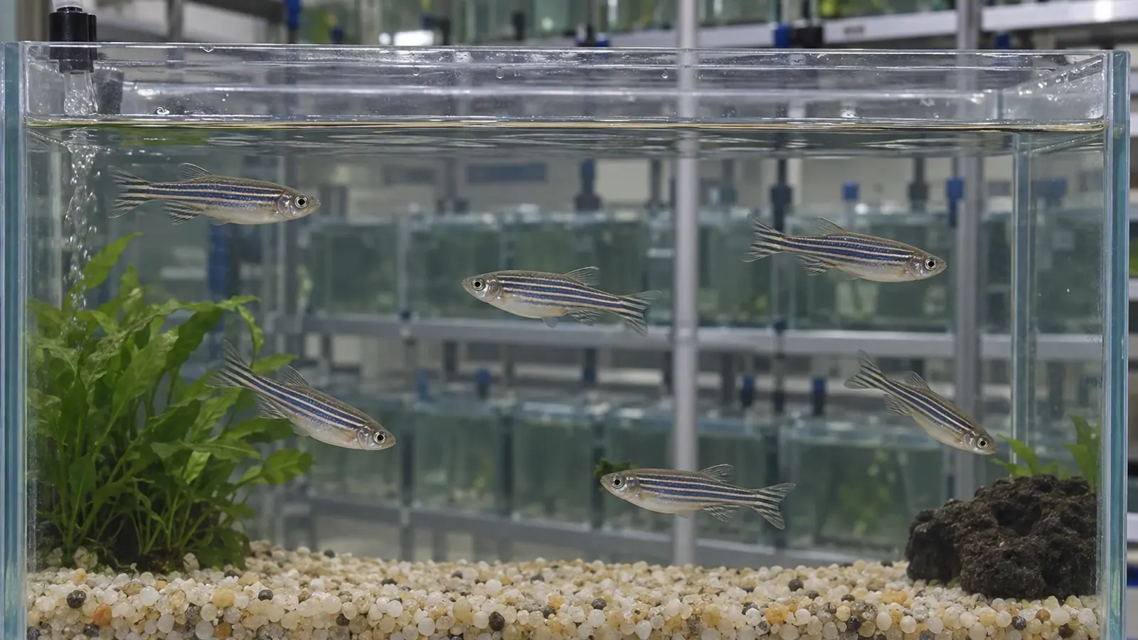
Zebrafish egg-associated inflammation: investigate first
A distended abdomen does not confirm egg retention. Zebrafish egg-associated inflammation requires a usable reproductive history and tissue diagnosis; the evidence does not establish a universal spawning schedule.


- Content type
- Practical guide
- Sector
- Research facilities
- Animal group
- Zebrafish
A swollen abdomen is an observation, not a diagnosis
During reproductive monitoring, a female zebrafish, Danio rerio, may develop a more prominent abdomen. That appearance does not establish egg retention or ovarian inflammation. Describe it alongside the fish’s feeding, swimming, previous appearance and spawning history. The first useful step is to keep what was observed separate from the proposed explanation.
The Zebrafish International Resource Center disease manual describes egg-associated inflammation, or EAI. Kent, Harper and Wolf’s 2012 review also discusses the condition with fibroplasia, referred to as EAIF. Together they provide a framework for investigation in a research facility. They do not establish a spawning frequency that will prevent every case.
What the evidence describes
EAI involves chronic inflammation associated with degenerating oocytes, and fibrous tissue may develop. Histopathology identifies the tissue changes. Abdominal shape alone cannot confirm the condition, and affected fish may lack obvious abdominal distension.
The sources discuss abnormal retention and insufficient opportunities to spawn as possible contributors. However, the precise cause remains unknown. Kent and colleagues note that inflammation and oocyte atresia can occur together without resolving which came first. The presence of degenerating eggs therefore does not automatically identify a scheduling failure.
The ZIRC manual recommends timely spawning opportunities. It also reports that manually expressing eggs has been attempted without reliable curative success. That observation is not an instruction to compress the abdomen of a suspected case. Any intervention should be assessed by the veterinarian in relation to the individual fish and the available evidence.
Reconstruct actual spawning events
A breeding attempt and a confirmed spawn need separate entries. For an identifiable female or group, record attempted pairings, observed eggs, partner changes, transfers and periods without spawning opportunities. A completed breeding task in a staff log does not necessarily mean that the intended biological event occurred.
Set those entries alongside known age, lineage and housing information. Make missing details explicit. An incomplete record cannot establish that a fish failed to spawn. Conversely, a recent pairing does not demonstrate normal ovarian emptying or exclude an underlying condition.
Traceability also helps assess the breeding programme. The facility should be able to explain why females are retained, how their condition is reviewed and what prompts reassessment. A written calendar helps staff organise work, but its existence alone does not establish that reproduction is successful for every fish.
When records are maintained by group rather than individual, keep that limitation visible. A successful group spawn cannot be assigned automatically to each female present. The available identification method should determine how specifically the history is interpreted.
Observe changes without unnecessary handling
A monitoring record can describe body outline, abdominal symmetry, swimming, food access and interactions. Repeated observations under comparable conditions are more interpretable than unplanned handling episodes. Staff should identify whether a change is recent, progressive or already documented.
Photographs taken from a consistent position can show the progression of external appearance. They cannot reveal internal tissue architecture. Distension remains a non-specific clinical sign. A rounded female may need investigation, but the image cannot name the disease or prove that every less rounded female is unaffected.
If the fish cannot feed, maintain position or reach necessary resources, prompt individual assessment is needed. Humane endpoints should follow the facility’s approved framework. This guide does not set an acceptable waiting period or create a universal clinical score.
Observers should record when they cannot assess an activity reliably. A fish hidden from the feeding view should not simply be marked as having refused food. That distinction avoids adding uncertainty to the clinical history while making the next observation more purposeful.
Preserve the differential diagnosis
The sources also describe other conditions affecting abdominal organs, including mycobacterial infections and masses. Keep alternative explanations open until appropriate examinations have been performed. Finding mycobacteria in some affected ovaries does not establish that they cause every instance of EAI.
Plan sampling with the diagnostic laboratory. Histology examines the architecture and distribution of lesions, whereas microbiological investigation answers a different question and may need different preservation. Discuss fixation before it prevents a useful additional test.
The submission should include animal identity, clinical observations, reproductive history and any treatment already given. A comparison involving other animals may be useful when justified by the veterinarian, but selecting every fish with a large abdomen is not, by itself, a diagnostic design. Sample selection should follow the question being asked.
Review prevention without inventing a fixed schedule
Appropriate spawning opportunities belong in the husbandry programme and should be reviewed against actual outcomes. Increasing the number of pairings automatically does not demonstrate effective prevention. Document changes and follow their consequences for welfare and reproductive performance.
The team can examine prolonged periods without reproductive opportunities, recurrent unsuccessful pairings and retained animals whose purpose or monitoring is unclear. These are management questions, not proven causes in every case. They help identify a specific adjustment whose effects can be assessed.
An ovarian disorder also matters when experiments measure fecundity or inflammation. Kent and colleagues identify possible interference with research interpretation. Do not silently exclude animals retrospectively or assign a lesion to an experimental treatment simply because health history is unavailable. Decisions on data handling need the study’s defined rules and a documented assessment.
The practical conclusion
EAI and EAIF are identified through tissue examination. Abdominal distension and an absence of recorded spawning can guide investigation, but they do not replace diagnosis. Before intervention, assemble a readable history, observe consistently and plan the diagnostic approach.
Vetofish’s diagnostic and analytical services support research facilities with observation criteria, sample planning and interpretation of findings. The purpose is to connect reproductive records, health and welfare, without assuming a cause or presenting an unvalidated manipulation as a cure.


